44 labeled diagram of a human cell
PDF Well Labeled Diagram Of Human Speech Organ May 7th, 2018 - This article explains the bone structure of the human body using a labeled skeletal system diagram and a simple memorize the labeled human skeleton diagram''Human Body Wikipedia May 7th, 2018 - The Study Of The Human Body Involves Anatomy The Body Is Also Host To About The Same Number Of Non Human Cells As Well As Multicellular ... Human Body Diagram - Bodytomy However, internally, the structure is far complex and intricate. Know that there are 11 organ systems of the body: Circulatory System, Respiratory System, Immune System, Skeletal System, Excretory System, Urinary System, Muscular System, Endocrine System, Digestive System, Nervous System, and Reproductive System.
Structure of Cell: Definition, Types, Diagram, Functions - Embibe What is a cell? Ans: The cell is the smallest, fundamental and functional unit that makes up all living beings including microorganisms, plants, animals and humans. Q.2. What are the five cell structures? Ans: A cell consists of many different structures that have definite shapes, structures, and functions of their own. Some of these structures ...

Labeled diagram of a human cell
Labeled Plant Cell With Diagrams | Science Trends The parts of a plant cell include the cell wall, the cell membrane, the cytoskeleton or cytoplasm, the nucleus, the Golgi body, the mitochondria, the peroxisome's, the vacuoles, ribosomes, and the endoplasmic reticulum. Parts Of A Plant Cell The Cell Wall Let's start from the outside and work our way inwards. Learn the parts of a cell with diagrams and cell quizzes Cell diagram unlabeled. It's time to label the cell yourself! As you fill in the cell structure worksheet, remember the functions of each part of the cell that you learned in the video. Doing this will help you to remember where each part is located. Click the links below to download the labeled and unlabeled eukaryotic cell diagrams. Cells Diagram | Science Illustration Solutions - Edrawsoft Cells Diagram. Cells are the basic building blocks of all living things. The human body is composed of trillions of cells. Cells have many parts, each with a different function. Some of these parts, called organelles, are specialized structures that perform certain tasks within the cell. Drawing cells diagram helps you better understand your ...
Labeled diagram of a human cell. Cell: Structure and Functions (With Diagram) - Biology Discussion Eukaryotic Cells: 1. Eukaryotes are sophisticated cells with a well defined nucleus and cell organelles. 2. The cells are comparatively larger in size (10-100 μm). 3. Unicellular to multicellular in nature and evolved ~1 billion years ago. 4. The cell membrane is semipermeable and flexible. 5. These cells reproduce both asexually and sexually. Diagram of human skin structure — Science Learning Hub Diagram of human skin structure. Add to collection. + Create new collection. Tweet. Rights: University of Waikato Published 1 February 2011 Size: 100 KB Referencing Hub media. The epidermis is a tough coating formed from overlapping layers of dead skin cells. Draw a labelled diagram of neuron and label any four different parts ... Draw a labelled diagram of neuron and label any four different parts explain Here is the description of human neuron along with the diagram of the neuron and their parts. The neuron is a specialized and individual cell, which is also known as the nerve cell. A group of neurons forms a nerve. 1. Anatomy and Physiology: Parts of a Human Cell - Visible Body To learn more about cells, check out our free Human Cell eBook! Cells can be divided into four groups: somatic, gamete, germ, and stem. Somatic cells are all the cells in the body that aren't sex cells, like blood cells, neurons, and osteocytes. Gametes are sex cells that join together during sexual reproduction. Germ cells produce gametes.
File:Diagram human cell nucleus.svg - Wikipedia A comprehensive diagram of a human cell nucleus. Date: 27 April 2006: Source: I did it myself with adobe ilustrator using the information found here , ,, and : Author: Mariana Ruiz LadyofHats: Permission (Reusing this file) Public domain Public domain false false: A Labeled Diagram of the Animal Cell and its Organelles A Labeled Diagram of the Animal Cell and its Organelles There are two types of cells - Prokaryotic and Eucaryotic. Eukaryotic cells are larger, more complex, and have evolved more recently than prokaryotes. Where, prokaryotes are just bacteria and archaea, eukaryotes are literally everything else. Labelled Diagram of Human Eye, Explanation and Function - VEDANTU There are two kinds of cells in the eye namely rods and cones. The basic functions of Rods and Cones are conscious light perception, color differentiation and depth perception. ... Labeled Diagram of Human Eye . The eyes of all mammals consist of a non-image-forming photosensitive ganglion within the retina which receives light, adjusts the ... Labeled Diagram of the Human Kidney - Bodytomy Labeled Diagram of the Human Kidney Quick Facts. Located in the abdominal cavity, kidneys are the most efficient filters. They are an important component of... Cross-section of a Human Kidney. The vital structural components of a kidney are enclosed in a smooth but tough fibrous... Structure of a ...
Plant and Animal Cell: Labeled Diagram, Structure, Function - Embibe Plant Cell: Plant cells are eukaryotic cells with a true nucleus along with specialized structures called organelles that carry out certain specific functions. Animal Cell: An animal cell is a type of eukaryotic cell that lacks a cell wall and has a true, membrane-bound nucleus along with other cellular organelles. Diagram of Plant and Animal Cell Types of Plant Cell- Definition, Structure, Functions, Labeled Diagram Palisade parenchyma cells are columnar elongated structured cells found in a variety of leaves, lying below the epidermal tissue. Palisades are closely linked cells in layers of mesophyll cells found in leaf cells. Ray parenchyma has both radial and horizontal arrangements majorly found within the stem wood of the plant. Human Cell Diagram, Parts, Pictures, Structure and Functions One of the few cells in the human body that lacks almost all organelles are the red blood cells. The main organelles are as follows : cell membrane; endoplasmic reticulum; Golgi apparatus; lysosomes; mitochondria; nucleus; perioxisomes; microfilaments and microtubules; Diagram of the human cell illustrating the different parts of the cell. Cell Membrane Cell Organelles Types (With Diagram) - Biology Discussion The following points highlight the ten main types of cell organelles present in the cell. The types are: 1. Nucleus 2. Plastids 3. Mitochondria 4. Endoplasmic Reticulum 5. Ribosomes 6. Lysosomes 7. Golgi Bodies 8.
Skin Model Project 65+ Super Ideas | Skin anatomy, Integumentary system ...
Labelled Diagram Of A Human Cell Bone Cell Labeled Diagram Animal Cell ... Labelled Diagram Of A Human Cell Bone Cell Labeled Diagram Animal Cell Free Printable To Label Find this Pin and more on Biology by Guinahi Douhe. Cell Structure Structure And Function Plasma Membrane Classroom Management Tips Label Image Animal Cell Application Letters Plant Cell Cell Wall More information ... More information
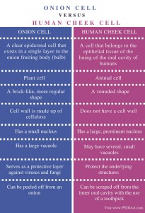
Difference Between Onion Cell and Human Cheek Cell - Pediaa.Com
Labeled Human Skeleton | Science Trends The labeled human skeleton system is comprised of 206 different bones of various sizes and shapes, all with the primary purpose of providing support, protection, and shape to the human body. ... Labeled Diagram Of The Human Skeleton. ... Bones also supply red and white blood cells that are made in the bone marrow, which is the material inside ...

Digestive System Diagram for Kids & Digestion Facts - InfoBarrel ...
Label a cell, Labeling parts of a cell, Cells Structures and Functions Start studying Label a cell, Labeling parts of a cell, Cells Structures and Functions. Learn vocabulary, terms, and more with flashcards, games, and other study tools.
Slide 35 - Cardiac Muscle - YouTube
A Labeled Diagram of the Plant Cell and Functions of its Organelles Unique to plant cells, the cell wall is a fairly rigid, protective wall that resists the strain of physical forces. The cell wall is mainly made up of cellulose fiber and it helps maintain the shape of the cell. Function: Maintains cell pressure and prevents over-expansion of the cell. Centrosome

Virus. Causes, symptoms, treatment Virus
Cell Membrane Diagram Labeled : Functions and Diagram Others include the following; a cell wall of peptidoglycan which keeps the final shape of the cell and it's made of polysaccharides and proteins. However, this cells have three distinctive shapes i.e spherical, rod shaped and spiral. The sole exception is Mycoplasma bacteria that have no cell wall and for that reason no particular shape.

Plant cell model 6th grade | Plant cell model, Cell model project ...
A Labelled Diagram Of Neuron with Detailed Explanations Diagram Of Neuron with Labels Here is the description of human neuron along with the diagram of the neuron and their parts. The neuron is a specialized and individual cell, which is also known as the nerve cell. A group of neurons forms a nerve.

Cross Section of Arm Through Musculospiral Nerve | ClipArt ETC
Animal Cells: Labelled Diagram, Definitions, and Structure Cell Organelles Plant Cells: Animal Cells: Cell wall: Present (made up of cellulose) Absent: Shape: Rectangular (fixed shape) Round (irregular shape) Vacuole: One, large central vacuole taking up to 90% of cell volume. One or more small vacuoles (much smaller than plant cells). Centrioles: Only present in lower plant forms (e.g. chlamydomonas)
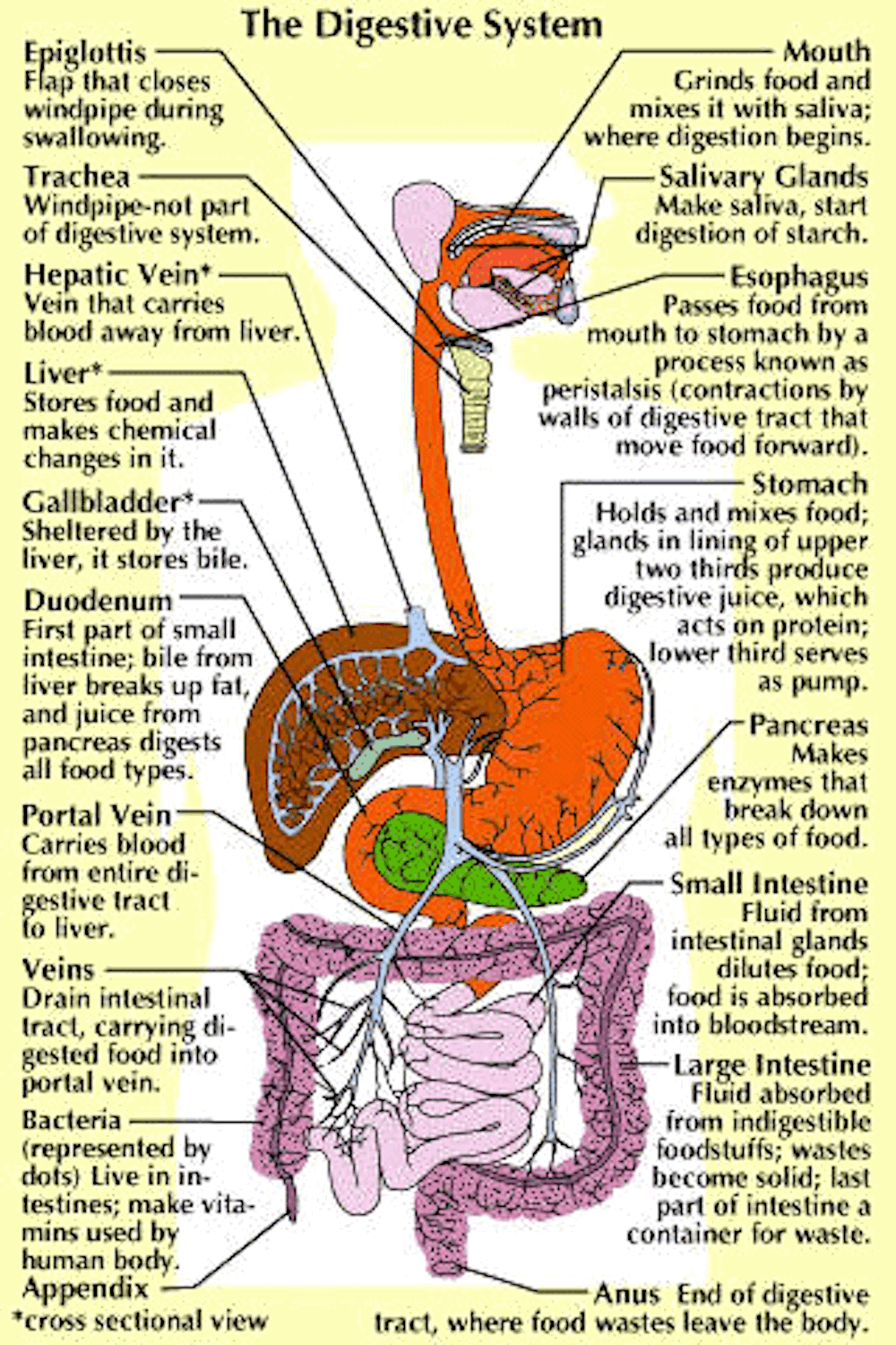
Parts and Function of Digestive System for Med School & Nursing ...
parts of a human cell | Diagram of the human cell illustrating the ... Human Drawing. Science Biology. Plant cells are covered by cell wall, it is a unique feature observed in plant cells. While the cell membrane acts as the outer layer in animal cells. The plant`s cell wall is made up of Cellulose. This is considered as major difference between plant cell and an animal cell.
ANAT2511 Introduction to Histology - Embryology
Structure and parts of a sperm cell - inviTRA This labelled diagram shows the structure of a sperm cellin detail, which has the following parts: Head With its spheric shape, it consists of a large nucleus, which at the same time contains an acrosome. The nucleus contains the genetic information and 23 chromosomes.
Post a Comment for "44 labeled diagram of a human cell"