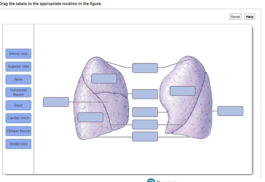
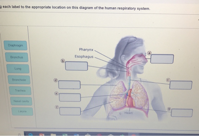
44 drag each label to the appropriate location on this diagram of the human respiratory system
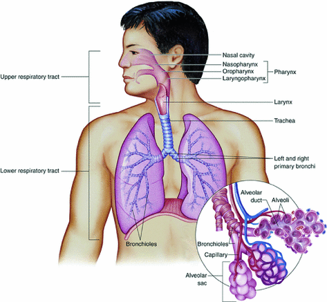
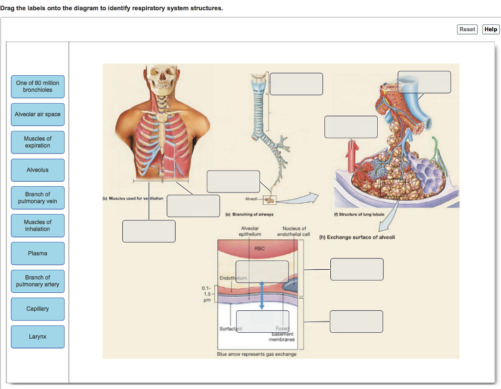
The Respiratory System - Diagram, Structure & Function The function of the human respiratory system is to transport air into the lungs and to facilitate the diffusion of oxygen into the bloodstream. It also receives waste Carbon Dioxide from the blood and exhales it. Here we explain the anatomy of the airways and how oxygen gets into the blood. The respiratory system organs are separated into the ... Drag each label to the appropriate location on the diagram. BioFlix Activity: Gas Exchange - The Respiratory System Lung Part A - The Respiratory System Drag Each Label To The Appropriate Location On This Diagram Of The Human Respiratory System. Reset Help Lary Pharynx Esophagus Trache Broncho Nasal Cave...
human basic organ anatomy location each quizlet system respiratory human appropriate drag label location diagram flashcards cardiovascular ch. Human Organ Diagram | Health Pictures | Human Organ Diagram, Human . human organs map organ body clipart diagram outline imgs internal clipartbest parts health. Labeled Diagram Of The Human Tongue - The Human Tongue Is A ...
:watermark(/images/watermark_only_sm.png,0,0,0):watermark(/images/logo_url_sm.png,-10,-10,0):format(jpeg)/images/anatomy_term/gracile-fasciculus-1/IgGqZ5xfdHvCthGXZdl4cw_gracile_fasciculus.png)
Drag each label to the appropriate location on this diagram of the human respiratory system
(Solved) - BioFlix Activity: Gas Exchange -- Oxygen Transport Drag each ... BioFlix Activity: Gas Exchange -- Oxygen Transport Drag each label to the appropriate location on the flowchart. 4 of 10 Reset Help Oxygen is carried through blood Oxygen diffuses from the alveoliinta Oxygen ditluses from the blood to the body's tissues Oxygen ones a red blood cell Oxygen binds to molecule of hemoglobin capilary Which of the following is part of the respiratory Part A - The respiratory system Drag each label to the appropriate location on this diagram of the human respiratory system. ANSWER: Correct HelpReset HelpResetAir travels down smaller and smaller bronchioles.Air enters through the nose or mouth. Air reaches small sacs called alveoli.Air travels down thetrachea and then enters the bronchi. Chapter 23 - Circulation & Respiration HW Flashcards - Quizlet The circulatory system of humans is composed of two loops: the systemic circulation, in which blood flows between the heart and lungs, and the pulmonary circulation, in which blood flows between the heart and the rest of the body. ... Drag each label to the appropriate location on this diagram of the human respiratory system. a. nasal cavity b ...
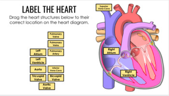
Drag each label to the appropriate location on this diagram of the human respiratory system. ear diagram without labels bones pelvic girdle diagram unlabeled labeling worksheet skeleton worksheeto via Drag Each Label To The Appropriate Location On This Diagram Of The atkinsjewelry.blogspot.com respiratory system label diagram human appropriate drag each location quiz Label The Ear Worksheets A& P II Chapter 16 Lab Flashcards | Quizlet Hormones help the body maintain homeostasis, or relatively constant internal conditions. Can you label the steps in a homeostasis pathway? To review the process of homeostasis, watch this BioFlix animation: Homeostasis: Regulating Blood Sugar. Drag each label to the appropriate location on the diagram of a homeostasis pathway. detailed skeletal system Digestive system diagram location human appendix facts digestion names teeth pain infobarrel untpikapps appropriate respiratory drag label each info body. Skeletal system part 1. Arts & crafts for kids : q-tip/cotton swab skeleton detailed skeletal system Diagram of Human Heart and Blood Circulation in It Four Chambers of the Heart and Blood Circulation. The shape of the human heart is like an upside-down pear, weighing between 7-15 ounces, and is little larger than the size of the fist. It is located between the lungs, in the middle of the chest, behind and slightly to the left of the breast bone. The heart, one of the most significant organs ...
respiratory system Flashcards | Chegg.com respiratory muscles Drag the appropriate labels to their respective targets. Label the figure that shows "INHALATION" and the figure that shows "EXHALATION" in targets (a) and (b). Then drag the other labels to the appropriate locations on the figures. During inhalation, the diaphragm and rib muscles contract. respiratory system gas transport worksheet label appropriate drag location each oxygen blood diffuses diagram carbon body tissues respiratory system dioxide lungs air enters into alveoli Human Respiratory System: MCQs Quiz - 4 human respiratory system quiz mcqs exchange questions lung its below question given gases shows correctly function breathing neet biology A&P2 Ch22 Respiratory System Homework Flashcards - Quizlet Respiratory system Drag each label to the appropriate location on this diagram of the human respiratory system. Path of air Drag each label to the appropriate location on the flowchart. Jane had been suffering through a severe cold and was complaining of a frontal headache and a dull, aching pain at the side of her face. Solved a each label to the appropriate location on this | Chegg.com Question: a each label to the appropriate location on this diagram of the human respiratory system. Diaphragm Pharynx Esophagus Bronchus Lung Bronchiole Trachea Nasal cavity Heart Larynx This problem has been solved! See the answer Show transcribed image text Expert Answer 100% (23 ratings)
Part a inhaling and exhaling part complete label the - Course Hero Part A Drag the terms on the left to the appropriate blanks on the right to complete the sentences. 1. The liquid portion of your blood is called plasma. 2. Erythrocytes, also called red blood cells, are packed with hemoglobin and transport oxygen to body tissues. 3. Leukocytes, also called white blood cells, function in fighting infections. MAP 6 - Respiratory System Flashcards - Quizlet Sort the correct pressures into the appropriate bins that represent tissue descriptions. Each bin should contain a value for PO2 and a value for Hb. answer in image: Resting Tissue = PO2 ~40 mm Hg, Hb ~75% O2 saturation. Metabolically Active tissue = PO2 ~20 mm Hg, Hb ~40% O2 saturation. Solved Part A - The respiratory system Drag each label to - Chegg a) Nasal cavity ( divided into 3 regions: I-Vestibular region has oil gland and tiny hair to filter air and trap dust paticles etc. II- Respiratory region has a layer of pseudostratified ciliated columnar epithelium which secretes mucuos. III- Olfact … View the full answer Answered: Drag the labels to the appropriate… | bartleby Drag the labels to the appropriate location in the figure. Reset Help Petrous part of Facial nervę Semicircular temporal (CN. ... pls help label the cat. A: The given diagram is the bony structure of a cat. The labelling is given in the following steps. ... The human respiratory system has a pair of lungs. Each lung is separated from the ...
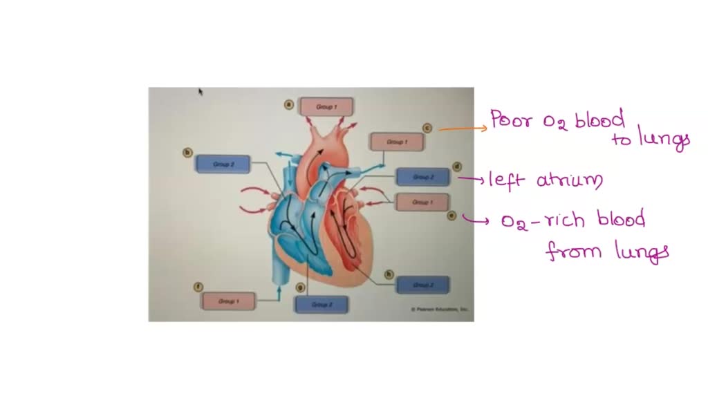
Drag Each Label to the Location of Each Structure Described In this interactive you can label parts of the human heart. The blood then returns to the left side of the heart where it is pumped to the rest of the body. Label each line on the pressure graph below as representing either the aorta left atrium or left ventricle.
Respiratory Homework - Subjecto.com Drag each label to the appropriate location on the flowchart. 1. Air enters through the nose or mouth 2. Air travels down the trachea and then enters the bronchi 3. Air travels down smaller and smaller bronchioles 4. Air reaches small sacs called alveoli. Drag each label to the appropriate location on this diagram of the human respiratory system.
Week 12: Respiration Flashcards - Quizlet A pneumothorax is the presence of air in the pleural cavity, which inhibits breathing. Labored breathing is called dyspnea. Small air passages less than 1 mm are called bronchioles. Higher than normal CO2 in the blood is called hypercapnia. diaphragm and external intercostals
From the capillaries of the abdominal organs and hind - Course Hero When air is inhaled, it follows a path through the respiratory system on its way to the alveoli. To review the human respiratory system, watch this BioFlix animation: Gas Exchange: The Path of Air into the Lungs. Part A - The respiratory system Drag each label to the appropriate location on this diagram of the human respiratory system.
(Solved) - BioFlix Activity: Gas Exchange - Transtutors BioFlix Activity: Gas Exchange ...
Respiratory System Anatomy, Diagram & Function | Healthline Respiratory. The respiratory system, which includes air passages, pulmonary vessels, the lungs, and breathing muscles, aids the body in the exchange of gases between the air and blood, and between ...
Ch-22_Gas Exchange.pdf - 5/6/2019 Ch-22_Gas Exchange Ch ... - Course Hero Part A - The respiratory system Drag each label to the appropriate location on this diagram of the human respiratory system. ANSWER: Help Reset Air enters through the nose or mouth. Air travels down the trachea and then enters the bronchi. Air travels down smaller and smaller bronchioles. Air reaches small sacs called alveoli.
Respiratory Homework Flashcards - Quizlet Drag each label to the appropriate location on the flowchart. 1. Oxygen diffuses from the alveoli into surrounding capillaries. 2. Oxygen enters a red blood cell. 3. Oxygen binds to a molecule of hemoglobin. 4. Oxygen is carried through blood vessels to a capillary. 5. Oxygen diffuses from the blood to the body's tissues During inhalation,
Answered: Drag the labels to the appropriate… | bartleby Science Anatomy and Physiology Q&A Library Drag the labels to the appropriate location in the figure. Reset Help Petrous Facial nervę part of Semicircular temporal (CN. VII) bone canals Facial nerve (N VI) Vęstibulocochlear nerve (CN. VIII) Vestibule Semicircular canals Bony labyrinth of internal ear Auditory tube Bony labyrinth of internal ear Petrous part of temporal bone Auditory tube ...
Chapter 23 - Circulation & Respiration HW Flashcards - Quizlet The circulatory system of humans is composed of two loops: the systemic circulation, in which blood flows between the heart and lungs, and the pulmonary circulation, in which blood flows between the heart and the rest of the body. ... Drag each label to the appropriate location on this diagram of the human respiratory system. a. nasal cavity b ...
Which of the following is part of the respiratory Part A - The respiratory system Drag each label to the appropriate location on this diagram of the human respiratory system. ANSWER: Correct HelpReset HelpResetAir travels down smaller and smaller bronchioles.Air enters through the nose or mouth. Air reaches small sacs called alveoli.Air travels down thetrachea and then enters the bronchi.
(Solved) - BioFlix Activity: Gas Exchange -- Oxygen Transport Drag each ... BioFlix Activity: Gas Exchange -- Oxygen Transport Drag each label to the appropriate location on the flowchart. 4 of 10 Reset Help Oxygen is carried through blood Oxygen diffuses from the alveoliinta Oxygen ditluses from the blood to the body's tissues Oxygen ones a red blood cell Oxygen binds to molecule of hemoglobin capilary









![MCQ] - Carefully study the diagram of the human respiratory ...](https://d1avenlh0i1xmr.cloudfront.net/e4eda889-b139-4abd-a93b-ff2178822628/human-respiratory-system---teachoo.png)








Post a Comment for "44 drag each label to the appropriate location on this diagram of the human respiratory system"