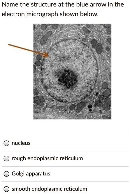
42 nucleus electron micrograph labelled
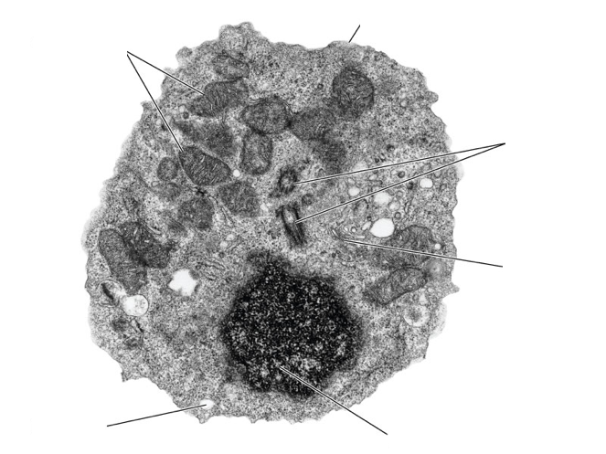
PDF Electron Micrographs (EMs) for laboratories in A215, Basic Human ... - IU Identify and be able to recognize: Label centrioleC The rod-like form of the centriole is seen in longitudinal (lengthwise) section near the right edge of Plate 12a. Note the l-micron scale to the lower left of the large, dark nucleus (N). CiteSeerX — nucleus by immunogold electron microscopy CiteSeerX - Document Details (Isaac Councill, Lee Giles, Pradeep Teregowda): The distribution of three adenovirus-encoded DNA replication proteins in the nucleus of human 293 cells was studied by immunogold electron microscopy. The infected nuclei contained four morphologically distinct inclusions. They were highly electron-dense granules (type I), compact fibrogranular masses of medium ...
Notes CELL STRUCTURE AND FUNCTION - National Institute of … z illustrate the structure of plant and animal cells by drawing labelled diagrams; z describe the structure and functions of plasma membrane, cell wall , endoplasmic reticulum (ER), cilia, flagella, nucleus, ribosomes, mitochondria, chloroplasts, golgi body, peroxisome, glyoxysome and lysosome; z describe the general importance of the cell molecules-water, mineral ions, …

Nucleus electron micrograph labelled
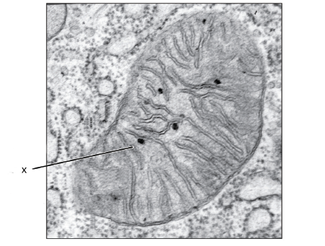
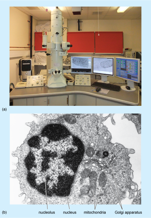
Electron Microscopy - University of Utah Plasma cell. Normal plasma cell with prominent cytoplasmic smooth endoplasmic reticulum. Macrophage. Normal macrophage with oblong nucleus, nucleolus, and cytoplasm with a variety of inclusions. Platelets. Normal platelets. Mitochondria. Happy mitochondria within a cell. Skeletal muscle. Dissecting the treatment-naive ecosystem of human melanoma … 07.07.2022 · Introduction. Melanoma brain metastases (MBMs) are the third most common cause of brain metastases after carcinomas of the lung and breast (Eichler et al., 2011) and lead to significant morbidity and mortality (Davies et al., 2011).While treatment with combination immune checkpoint blockade can be effective in patients with MBM (Tawbi et al., 2018), many … Plant Cell Nucleus Electron Micrograph - Dannie Vanlith When observed under the electron microscope, the nucleolus can be seen to consist of three distinguishable regions: In electron micrographs, centrioles appear as cylindrical structures which occur in pairs lying at right angles to each other (figs. Animal cell electron micrograph labelling. The nucleus controls the structure of the cell by transcribing dna which encodes for structural proteins such as actin plant cells generally have one large vacuole that takes up most of the cell's volume.
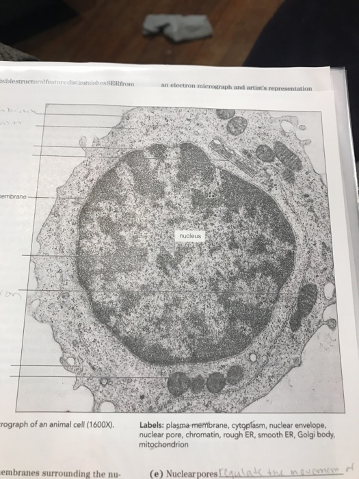
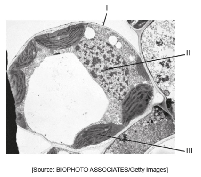
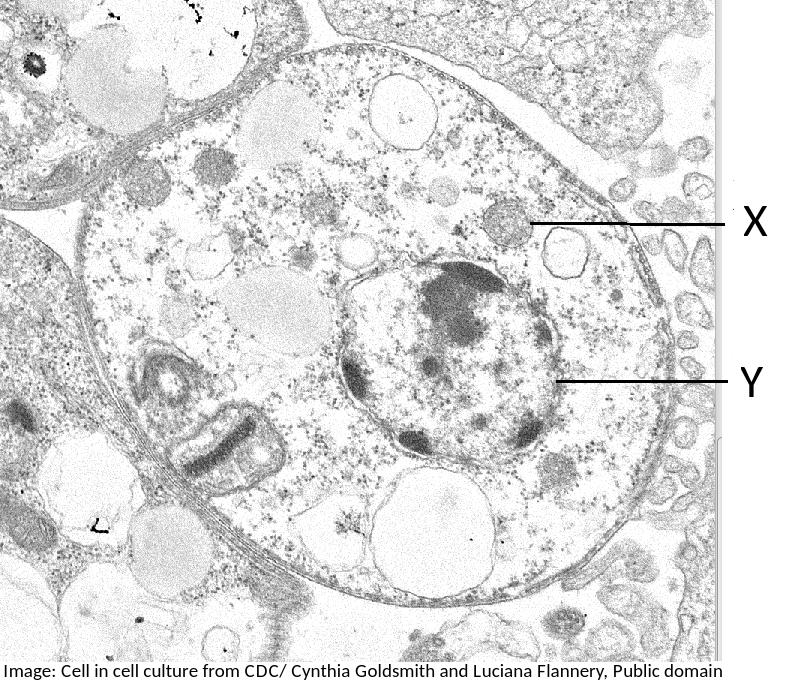
Nucleus electron micrograph labelled. Architecture and self-assembly of the jumbo bacteriophage … Aug 03, 2022 · The nucleus-like compartment formed in bacteria during infection by jumbo phage 201phi2-1 is composed of the bacteriophage protein chimallin, which can self-assemble into closed compartments in vitro. animal cell under electron microscope labelled - Be A Terrific Memoir ... Tuesday April 20th 2021. Animal Cell Diagram Under Microscope Labeled. Here is an electron micrograph of an animal cell with the labels superimposed. An animal cell represents an eukaryotic cell in which true nucleus and other membrane-bound organelles such as mitochondria Golgi bodies and lysosomes are present. Function cell does in the body. Glossary of botanical terms - Wikipedia pl. apices The tip; the point furthest from the point of attachment. aphananthous (of flowers) Inconspicuous or unshowy, as opposed to phaneranthous or showy. aphlebia pl. aphlebiae Imperfect or irregular leaf endings commonly found on ferns and fossils of ferns from the Carboniferous Period. aphyllous Leafless; having no leaves. apical At or on the apex of a … plant cell label electron micrograph Diagram | Quizlet Start studying plant cell label electron micrograph. Learn vocabulary, terms, and more with flashcards, games, and other study tools.
Ultrastructure and nuclear architecture of telomeric chromatin revealed ... Superimposition of eGFP fluorescence signals and the corresponding EM micrographs from eGFP-APEX2-labelled TRF2 in MEFs, TRF1 in MEFs and TRF1 in U2OS cells demonstrate that telomeres labelled by the APEX2 probes are more electron dense than other chromatin regions in the nucleus (Figure (Figure1C 1C - G). Cell Nucleus - function, structure, and under a microscope The nucleus is a double-layer membrane organelle. It consists of the nuclear envelope, DNA (chromatin), nucleolus, nucleoplasm, and the nuclear matrix. The main function of the nucleus is to control cell activities and carry genetic information to pass to the next generation. A eukaryotic cell typically has only one nucleus. Nucleus - Electron Micrograph - University of Tulsa Slide 5 of 36 animal cell electron micrograph labelling Diagram | Quizlet Start studying animal cell electron micrograph labelling. Learn vocabulary, terms, and more with flashcards, games, and other study tools.
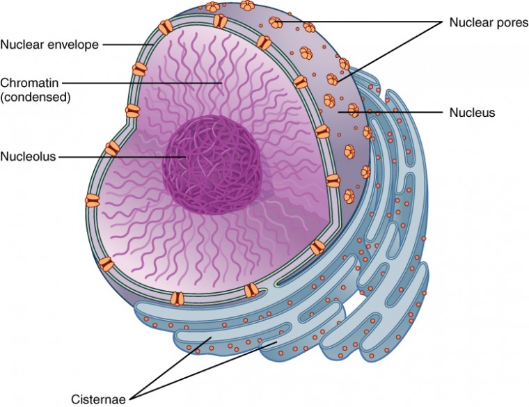
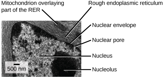
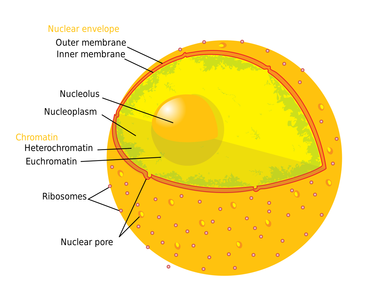
Nucleus: Definition, Structure, Functions - Biology Learner A typical nucleus has four parts seen in the electron microscope: Nuclear membrane; Nucleolus; Nucleoplasm or Nuclear sap; Nuclear reticulum; Figure: Labelled diagram of Nucleus and its different parts . Nuclear Membrane. ... The electron micrograph and immunocytological techniques show that three distinct regions are observed in the nucleolus. Label This Transmission Electron Micrograph - Kaiden Brown Label the transmission electron micrograph of the nucleus. Molecular labeling for correlative microscopy: Subset of labeled images and transfer labels to the entire image corpus. Label this transmission electron micrograph of relaxed sarcomeres by clicking and dragging the labels to the correct location . Electron Micrographs of Cell Organelles | Zoology - Biology Discussion The Electron Micrograph of Nucleus: This is an electron micrograph of nucleus. (Fig. 17 & 18): (1) Nucleus was discovered by Brown (1831). (2) It is a characteristic entity of almost all eukaryotic cells except mammalian RBCs. (3) The nucleus is generally one but may also be two, four or many. Angiosperm Life Cycle - Digital Atlas of Ancient Life 09.08.2019 · Overview. The angiosperm life cycle, in many ways, follows the basic life cycle pattern for land plants (embryophytes), with modifications characteristic of the seed plant habit (read more here).). As in other seed plants, the microgametophyte (male, or sperm-producing gametophyte) is highly simplified and called a pollen grain.The megagametophyte (female, or …
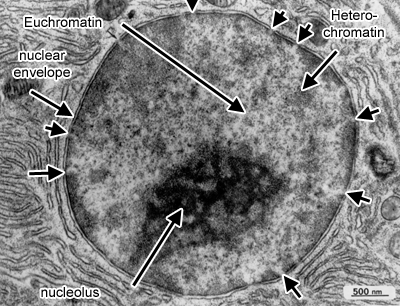
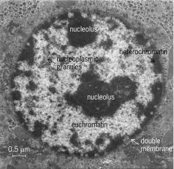
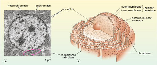
Electron Micrographs - University of Oklahoma Health Sciences Center Below is a collection of electron micrographs with labelled subcellular structures that you should be able to identify. Also, be sure to observe any electron micrographs which are made available in the laboratory by the instructor. ... Figure 1 Micrograph of a nucleus. 1. Heterochromatin 2. Euchromatin 3. Nucleolus 4. Nucleolar associated ...
Labeled Diagram Of Cell Membrane : Electron Micrograph Electron Micrograph from In other words, a diagram of the membrane (like the one below) is just a snapshot of a dynamic process in which phospholipids and proteins are continually . Some of the major parts of the plasma membrane are : How do we know what we know about cells? 1)cell membrane 2)vacuole 3)nucleus 4)endoplasmic reticulum 5)mitochondria 6)golgi body.
PDF Identifying Organelles from an Electron Micrograph - Ms JMO's Biology ... The electron micrograph displayed below illustrates many of the plant cell characteristics discussed The cell wall, large central vacuole and chloroplasts are clearly visible Also visible is the clearly defined nucleus containing chromatin
Electron Micrograph of a Lymphocyte - Netter Images Electron Micrograph of a Lymphocyte. Variant Image ID: 18920. Add to Lightbox. Email this page. Link this page. Print. Please describe! how you will use this image and then you will be able to add this image to your shopping basket.
Animal Cell Electron Microscope Labelled - Q14 Draw a large diagram of ... Electron microscopes use accelerated electron beams (as opposed to visible light in a light microscope) to create images of magnification as here is an electron micrograph of an animal cell with the labels superimposed: (i) name the parts labelled as 1 to 10.
Solved Please label the electron micrograph to assess your | Chegg.com Expert Answer. 100% (3 ratings) 1 ) Nuclear envelo …. View the full answer. Transcribed image text: Please label the electron micrograph to assess your knowledge of the structure and function of a cell's nucleus nuclear pore endoplasma reticulum chromatin nucleolus nuclear envelope. Previous question Next question.
(PDF) [Easterling, Kenneth E.; Porter, Phase ... - Academia.edu Enter the email address you signed up with and we'll email you a reset link.
Nanoscale imaging of phonon dynamics by electron microscopy 08.06.2022 · The control of phonon propagation and thermal conductivity of materials by nanoscale structural engineering is exceedingly important for the development and improvement of nanotransistors, thermal ...
Deformation twinning - ScienceDirect 01.01.1995 · The concepts of twinning shears and twinning modes are introduced. The early attempts to predict these features are presented. This is followed by a detailed discussion of the formal theories of Bilby and Crocker and Bevis and Crocker for predicting these elements.
Solved Label the transmission electron micrograph of the - Chegg 100% (23 ratings) Transcribed image text: Label the transmission electron micrograph of the nucleus. Nuclear envelope Nucleolus Nucleus Heterochromatin Reset Zoom.
Tomography of the cell nucleus using confocal microscopy and medium ... The migration of BrUTP-labeled rRNAs was observed in α-amanitin (an inhibitor of RNA polymerase II)-treated cells which were pulse-labeled for 15 min and then chased for 2, 15, 30 and 90 min ().Visualization of the same cell firstly by phase-contrast microscopy and secondly by confocal microscopy showed that rRNA synthesis takes place in tiny structures within nucleoli.
Electron Micrograph of a Neutrophil - Netter Images Electron Micrograph of a Neutrophil. Variant Image ID: 13578. Add to Lightbox. Email this page. Link this page. Print. Please describe! how you will use this image and then you will be able to add this image to your shopping basket.
Cambridge International AS and A Level Biology ... - Academia.edu BIO1: Maintaining a Balance 1. Most organisms are active in a limited temperature range IDENTIFY THE ROLE OF ENZYMES IN METABOLISM, DESCRIBE THEIR CHEMICAL COMPOSITION AND USE A SIMPLE MODEL TO DESCRIBE THEIR SPECIFICITY ON SUBSTRATES
[Immune electron microscope determination of the localization of ... The number of particles observed over diffuse chromatin equals to 50-80% against the label in fibroblast cytoplasm. In contrast, the label used to be absent over the E. coli nucleoid. The presence of TRS in the fibroblast nucleus may evidence in favour of a possible regulatory role of TRS in eukaryots.
Label the transmission electron micrograph of the nucleus. - Transtutors Posted 3 days ago. View Answer . Q: 1. The following data refers to the demand for money (M) and the rate of interest (R) in for eight different economics: M (In billions) 56 50 46 30 20 35 37 61 R% 6.3 4.6 5.1 7.3 8.9 5.3 6.7 3.5 a. Assuming a relationship , obtain the OLS estimators...
Electron micrographs of SPIO-labeled MSCs. A, Cell nucleus (N) and ... Download scientific diagram | Electron micrographs of SPIO-labeled MSCs. A, Cell nucleus (N) and endosomal vesicles containing SPIO particles. (Original magnification, ϫ 6600.) B, Iron-loaded ...
Neuron under Microscope with Labeled Diagram - AnatomyLearner The postsynaptic membrane is an electron density that similar to the presynaptic density. This density reflects the high protein content of the synaptic membrane. Neuron under microscope labelled diagram. Throughout this article, you got the different neurons labelled diagrams.
The Cell: The Histology Guide - University of Leeds An electron micrograph of a nucleus Types of Nucleus Cells are normally diploid - this means that they have a pair - two sets of homologous chromosomes, and hence two copies of each gene or genetic locus. However, cells can be haploid, polyploid or aneuploid. Haploid: only has one set of chromosomes - i.e. in a sperm or oocyte.
Nucleus reticularis thalami connections with the mediodorsal ... - PubMed Postembedding immunocytochemistry for GABA was performed on the tissue containing anterograde WGA-HRP label for identification of NRT boutons under electron microscope. The double-labeled boutons were of small to medium size, contained a large number of pleomorphic vesicles, few mitochondria, and formed multiple symmetric synaptic contacts.
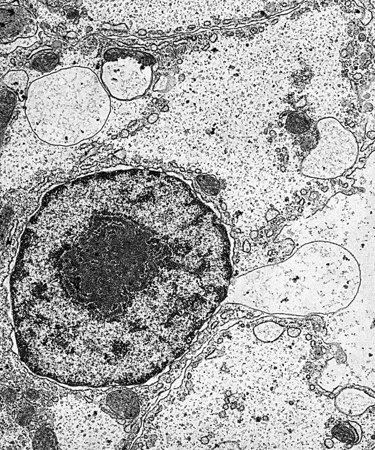
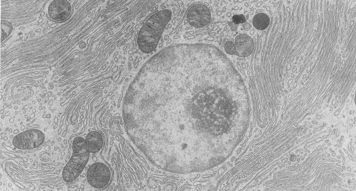
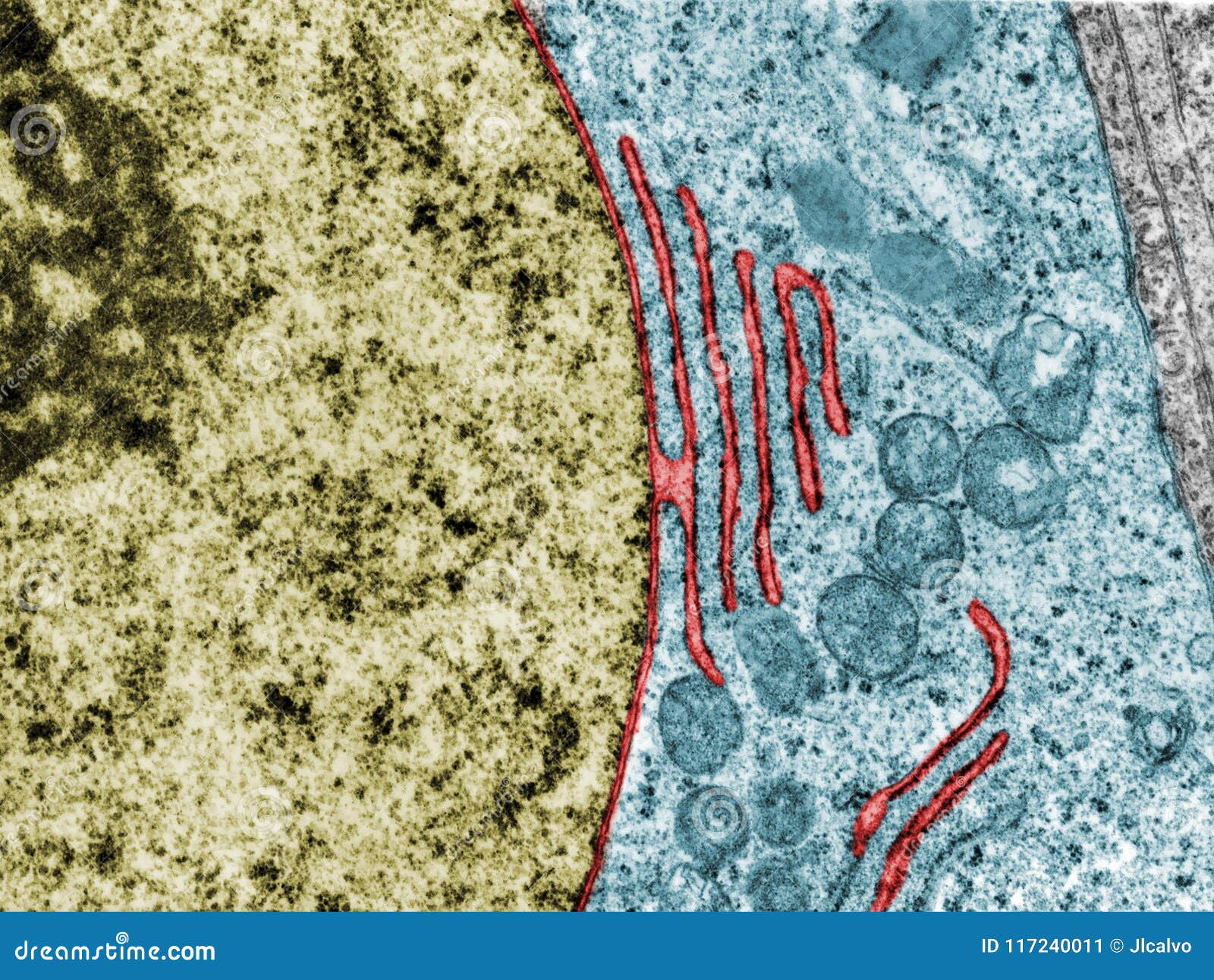
Cambridge International AS & A Level Biology Coursebook … Figure 1.23: Transmission electron micrograph (TEM) of a nucleus. This is the nucleus of a cell from the pancreas of a bat (×11000). The circular nucleus is surrounded by a double-layered nuclear envelope containing nuclear pores. The nucleolus is more darkly stained. Rough ER is visible in the surrounding cytoplasm. es w ie nuclear pore ...
Virtual EM Micrograph List | histology - University of Michigan 021. Plasma Cell: This electron micrograph shows a typical secretory cell, a plasma cell, which secretes immunoglobulin protein. Many of the major types of cellular organelles are visible in this image. In the nucleus, areas of euchromatin and heterochromatin can easily be identified. Virtual Slide.
Plant Cell Nucleus Electron Micrograph - Dannie Vanlith When observed under the electron microscope, the nucleolus can be seen to consist of three distinguishable regions: In electron micrographs, centrioles appear as cylindrical structures which occur in pairs lying at right angles to each other (figs. Animal cell electron micrograph labelling. The nucleus controls the structure of the cell by transcribing dna which encodes for structural proteins such as actin plant cells generally have one large vacuole that takes up most of the cell's volume.
Dissecting the treatment-naive ecosystem of human melanoma … 07.07.2022 · Introduction. Melanoma brain metastases (MBMs) are the third most common cause of brain metastases after carcinomas of the lung and breast (Eichler et al., 2011) and lead to significant morbidity and mortality (Davies et al., 2011).While treatment with combination immune checkpoint blockade can be effective in patients with MBM (Tawbi et al., 2018), many …
Electron Microscopy - University of Utah Plasma cell. Normal plasma cell with prominent cytoplasmic smooth endoplasmic reticulum. Macrophage. Normal macrophage with oblong nucleus, nucleolus, and cytoplasm with a variety of inclusions. Platelets. Normal platelets. Mitochondria. Happy mitochondria within a cell. Skeletal muscle.
















Post a Comment for "42 nucleus electron micrograph labelled"