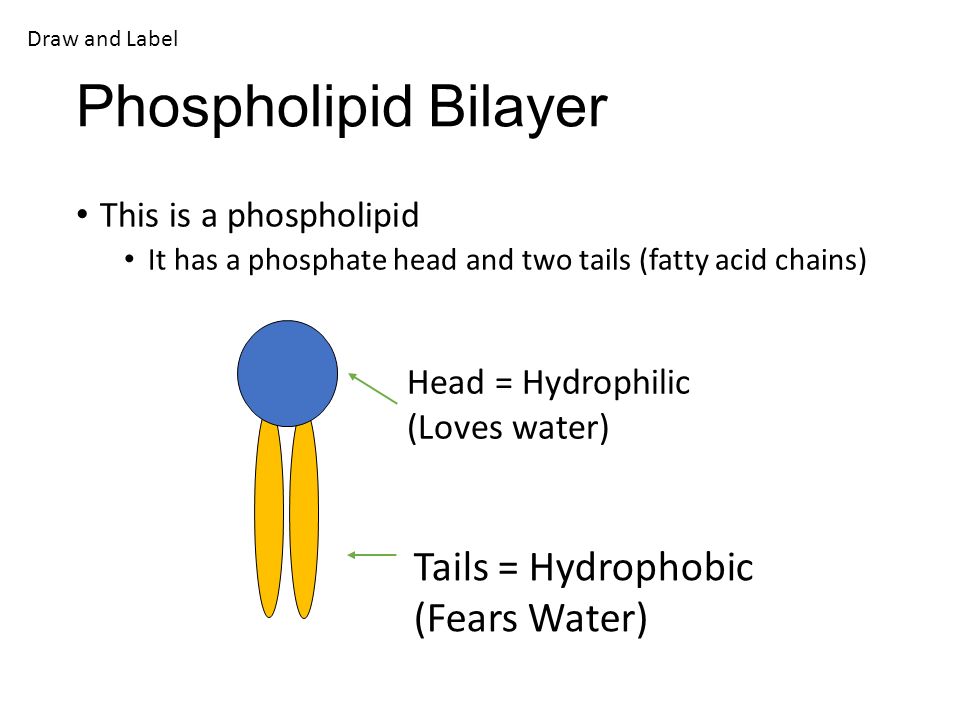
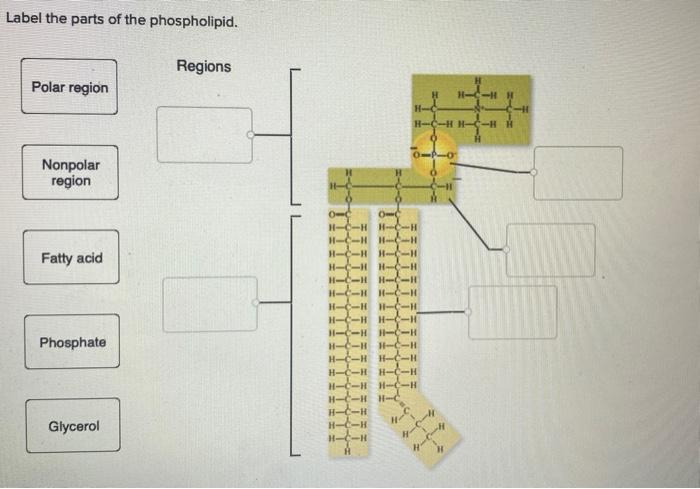
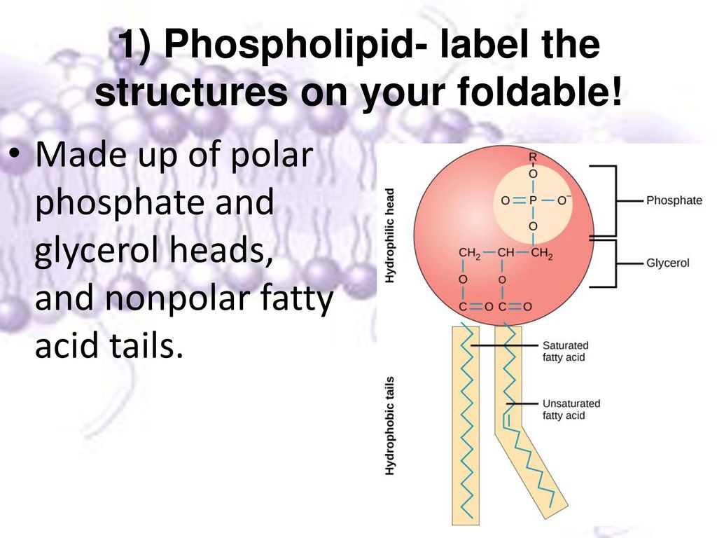
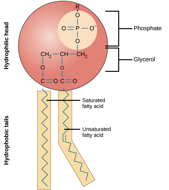
43 draw a phospholipid and label its parts
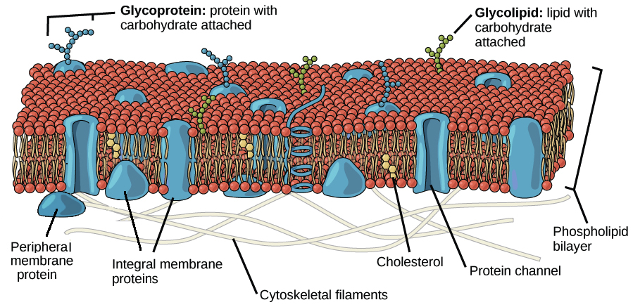
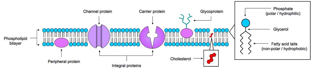
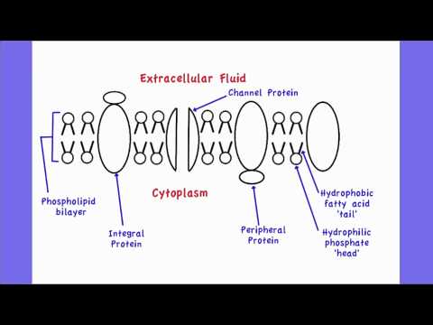
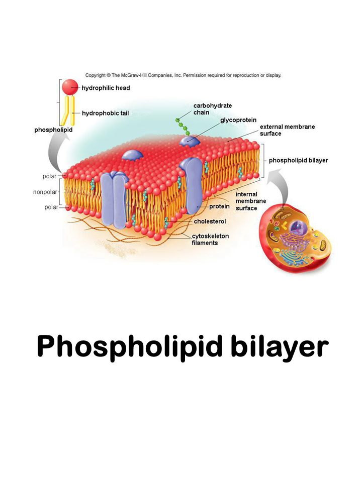
Cell (biology) - Wikipedia Hence, the layer is called a phospholipid bilayer, or sometimes a fluid mosaic membrane. Embedded within this membrane is a macromolecular structure called the porosome the universal secretory portal in cells and a variety of protein molecules that act as channels and pumps that move different molecules into and out of the cell. [4] The human cell is composed of three basic components: the ... According to the fluid-mosaic model of the plasma membrane, a.protein and phospholipids form a regular, repeating structure. b.the membrane is a rigid structure. c.phospholipids form a double layer, with the polar parts facing each other. d.proteins are free to move within a double layer of phospholipids.
Construction of the Cell Membrane - Wisc-Online OER In this interactive object, learners read the definitions of the parts of a cell and assemble a basic eukaryotic cell in a drag and drop exercise. A matching quiz involving cell terms and their definitions completes the activity.

Draw a phospholipid and label its parts
Answered: In the lungs, there are steep… | bartleby In the lungs, there are steep concentration gradients for oxygen and carbon dioxide molecules such that large numbers of these molecules move across the plasma membrane of the cells that line the lungs. Exosome: A Review of Its Classification, Isolation Techniques ... Sep 22, 2020 · Introduction. Exosomes, with a diameter of about 40–100nm, are biological nanoscale spherical lipid bilayer vesicles secreted by cells, floating at a density of 1.13–1.19 g ∙ mL −1 in a sucrose density gradient solution. 1–5 In 1981, Trams et al 6 collectively referred to plasma membrane-derived vesicles as exosomes and first proposed the concept of “exosomes”, which was regarded ... Maharashtra Board Class 10 Science Solutions Part 2 Chapter 2 ... Jan 13, 2021 · Draw a neat diagram of the structure of chromosome and label the parts: (a) Centromere (b) p-arm (March 2019) Answer: Question 2. Sketch and label the diagram to show ATP – the energy currency of the cell. Answer: Question 3. Mitochondria and Krebs cycle: Answer: (a) Which co-enzymes are shown in the diagram? Answer:
Draw a phospholipid and label its parts. (PDF) Cambridge International AS and A Level Biology ... BIO1: Maintaining a Balance 1. Most organisms are active in a limited temperature range IDENTIFY THE ROLE OF ENZYMES IN METABOLISM, DESCRIBE THEIR CHEMICAL COMPOSITION AND USE A SIMPLE MODEL TO DESCRIBE THEIR SPECIFICITY ON SUBSTRATES Maharashtra Board Class 10 Science Solutions Part 2 Chapter 2 ... Jan 13, 2021 · Draw a neat diagram of the structure of chromosome and label the parts: (a) Centromere (b) p-arm (March 2019) Answer: Question 2. Sketch and label the diagram to show ATP – the energy currency of the cell. Answer: Question 3. Mitochondria and Krebs cycle: Answer: (a) Which co-enzymes are shown in the diagram? Answer: Exosome: A Review of Its Classification, Isolation Techniques ... Sep 22, 2020 · Introduction. Exosomes, with a diameter of about 40–100nm, are biological nanoscale spherical lipid bilayer vesicles secreted by cells, floating at a density of 1.13–1.19 g ∙ mL −1 in a sucrose density gradient solution. 1–5 In 1981, Trams et al 6 collectively referred to plasma membrane-derived vesicles as exosomes and first proposed the concept of “exosomes”, which was regarded ... Answered: In the lungs, there are steep… | bartleby In the lungs, there are steep concentration gradients for oxygen and carbon dioxide molecules such that large numbers of these molecules move across the plasma membrane of the cells that line the lungs.
Materials move across the Cell's membrane” - ppt download

Modulation of lipid biosynthesis by stress in diatoms ...
Structure of the plasma membrane (article) | Khan Academy

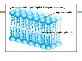
The Phospholipid molecule Hydrophilic – “water loving ...

Phospholipid Bilayer ( Read ) | Biology | CK-12 Foundation

Phospholipids (1.2.3) | AQA A Level Biology Revision Notes ...
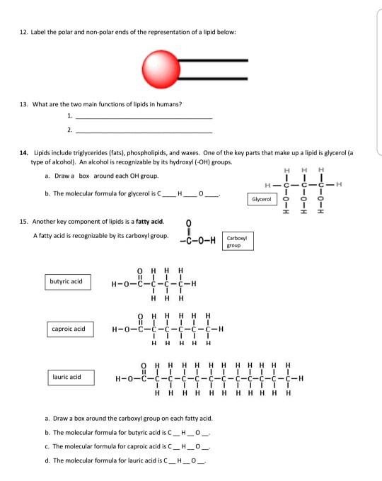
Chemistry of Lipids
13 List the 4 steps of the technological

Which cell organelle packages materials coming from the ...
Solved] Draw and label diagram of a cell membrane, showing ...

Fluid mosaic model: cell membranes article (article) | Khan ...
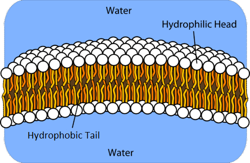
14.3: Phospholipids in Cell Membranes - Chemistry LibreTexts

How to Draw a Phospholipid Bilayer - YouTube

Schematic drawing of a squamous cell carcinoma denoting the ...
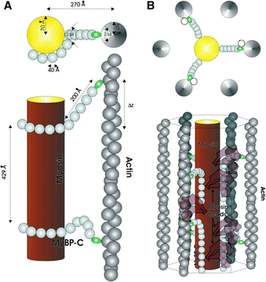
Myosin binding protein C: implications for signal ...
The Cell Membrane Write. Cell Membrane The membrane of the ...
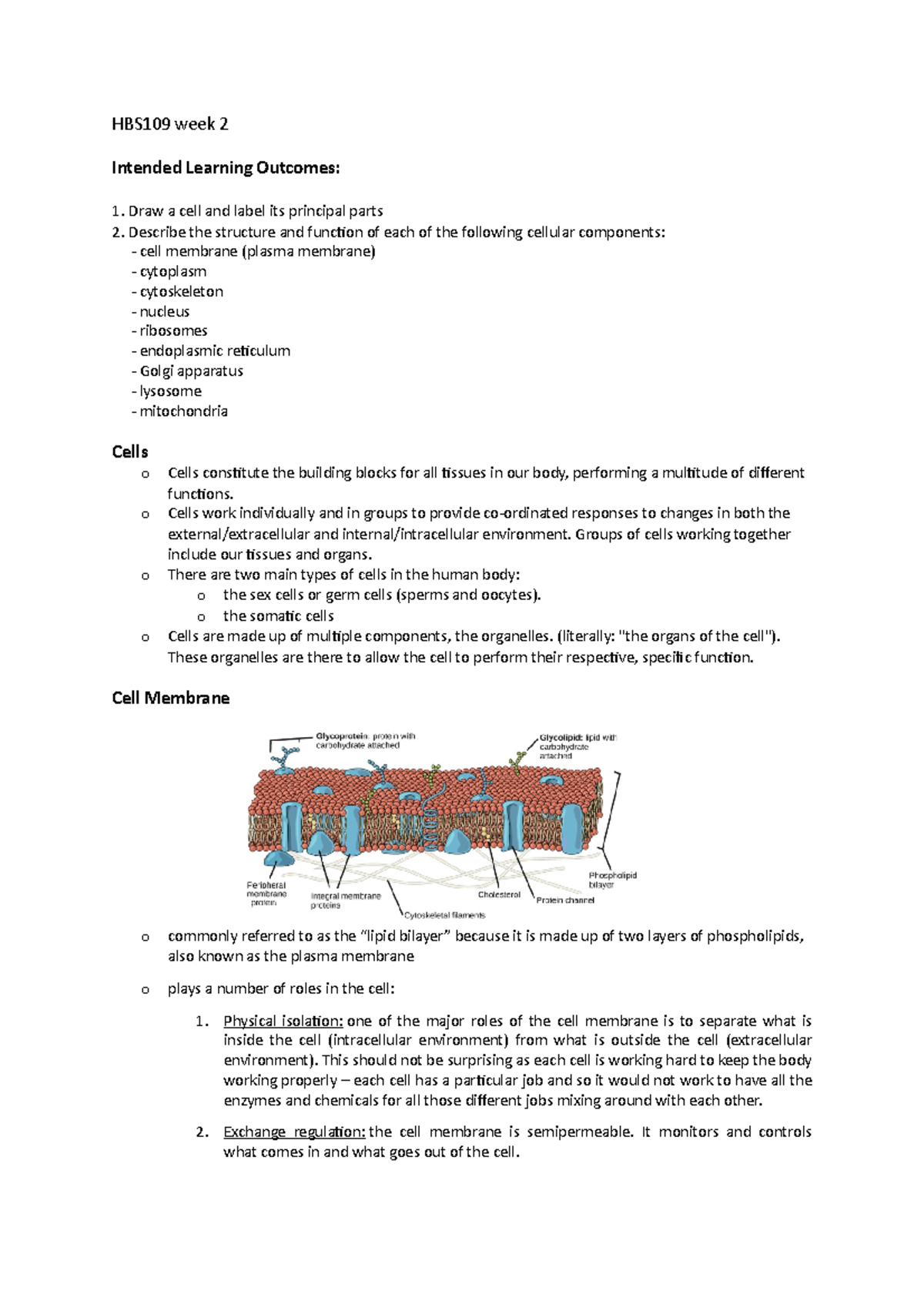
HBS109 week 2 notes - HBS109 week 2 Intended Learning ...

Photoactivatable Lipid Probes for Studying Biomembranes by ...
Solved Name: Pd: Lipids Worksheet 1. Are lipids organic ...
:max_bytes(150000):strip_icc()/cell-membrane-373364_final-5b5f300546e0fb008271ce52.png)
Cell Membrane Function and Structure
II. TINJAUAN PUSTAKA 2.1. Tanaman Eucalyptus sp. Eucalyptus ...

Biology, The Chemistry of Life, Biological Macromolecules ...
2.4 Membranes | BioNinja

Cell membrane Diagram | Quizlet
The Structure and Function of the Plasma Membrane
Solved Build a cell membrane Use the labels to draw and ...

Fluid mosaic model: cell membranes article (article) | Khan ...
Enzyme review questions What is an enzyme What

Phospholipid structure (video) | Khan Academy

Label the Phospholipid Bilayer Diagram | Quizlet
2.4.1 Draw and label a diagram to show the structure of membranes
Structure and function of connective tissue (Chapter 20 ...
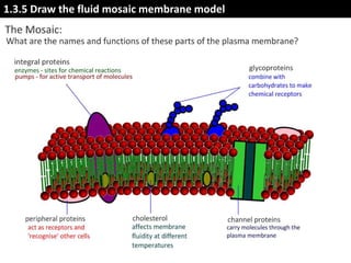
IB Biology 1.3 Slides: Membrane Structure
Phospholipid bilayer. Prepare to draw key components to help ...
Sensors | Free Full-Text | Spectrum Analysis of Gravity Waves ...
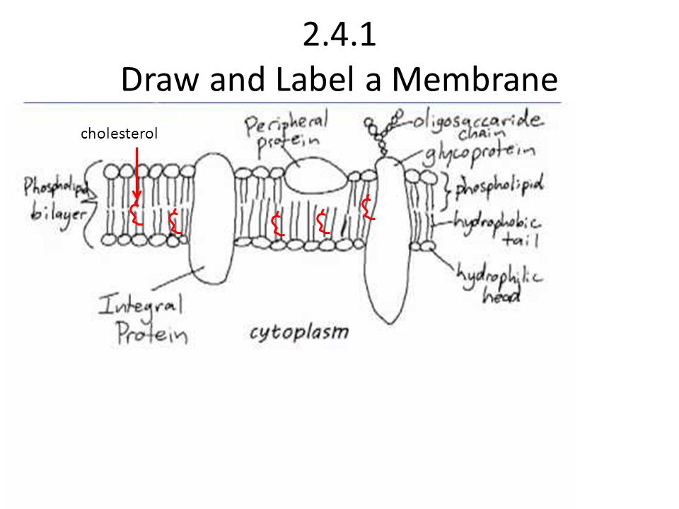
Topic 2.4 MEMBRANES Draw and Label a Membrane cholesterol ...

Chemical Structure of Lipids — Overview & Types - Expii
The Cell Membrane Biology Honors. - ppt download

Draw a well labelled diagram of animal cell and mention one ...
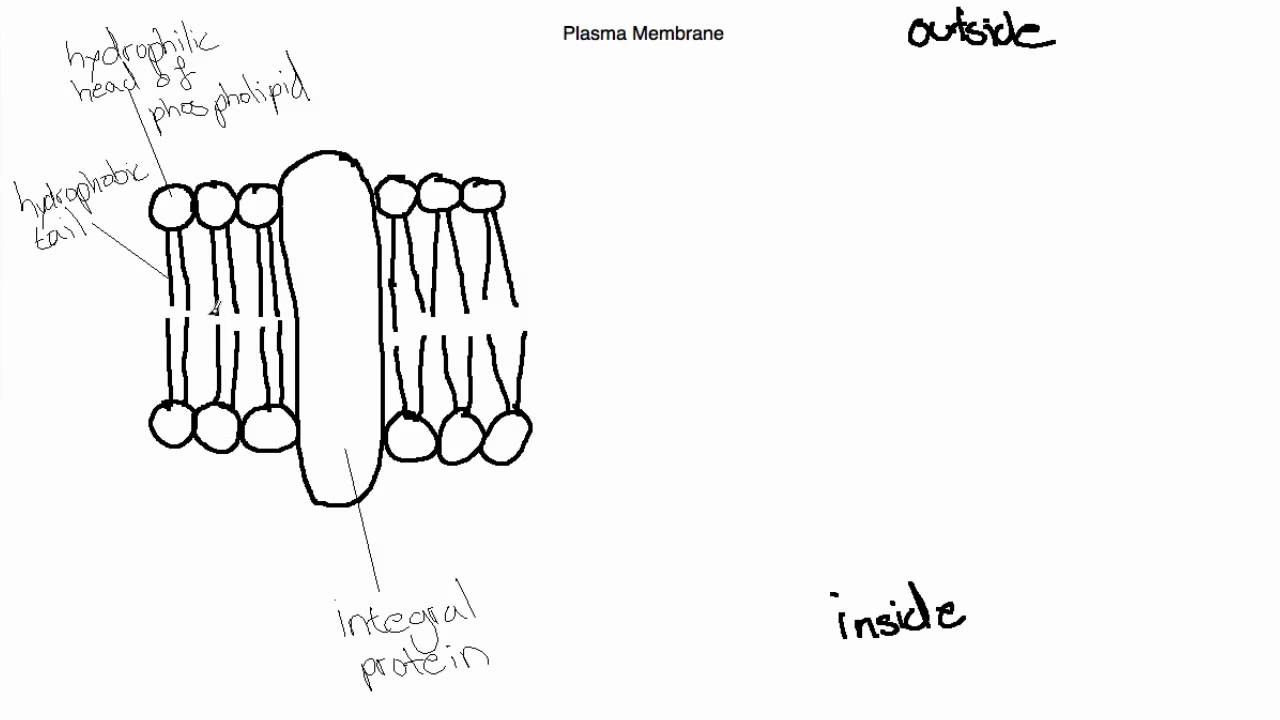
IB Biology Topic 2.4.1 Draw and Label the Plasma Membrane
Structure of the plasma membrane (article) | Khan Academy

Simple Lipid - an overview | ScienceDirect Topics

September 16th, 2019 Come in and grab your papers, journal ...
Post a Comment for "43 draw a phospholipid and label its parts"