44 diagram of neuron with labels
We found 13 reviewed resources for label a neuron diagram - Lesson Planet In this neuron worksheet, 7th graders study the definitions for the neuron and then label the neuron diagram with the terms. Get Free Access See Review + Lesson Planet: Curated OER. The Nervous System For Students 9th - 12th. Human biology beginners label colorful diagrams of the neuron, the reflex pathway, and the brain. They list steps in the ... A Labelled Diagram of Neuron with Detailed decription - Collegedunia Ans. Neurons can be classified as follows based on their polarity and the number of axons and dendrites: Unipolar: Only one structure extends from the cell body, or soma, of these neurons. Bipolar: One axon and one dendrite extend from the soma of these neurons. Multipolar Neurons: These neurons have a single axon and many dendrites. Purkinje cells, Anterior horn cells, and other cells are examples.
A Guide to Understand Neuron with Neuron Diagram 3. How to Draw a Neuron Diagram To learn about the structure of the neurons, the students can use a neuron labeled diagram. The students may follow these steps to make their neuron diagram, but the process is complex: 3.1 How to Draw a Neuron Diagram from Sketch Step 1: First, the students need to draw a circle. Based on it, they need to draw a star-like shape.

Diagram of neuron with labels
Diagram Quiz on Neuron Structure and Function (Labeling Quiz) Diagram Quiz on DNA replication. 1. Identify the cell type in the above figure. 2. In the figure, labeled '1' receives impulses from adjacent neuron. It is called the. 3. In the figure, labeled '2' is the short filaments from the cell body that carries impulses from dendrites to the cell body which is the. 4. byjus.com › biology › skin-diagramSkin Diagram with Detailed Illustrations and Clear Labels - BYJUS Skin Diagram The largest organ in the human body is the skin, covering a total area of about 1.8 square meters. The skin is tasked with protecting our body from external elements as well as microbes. Labeled Neuron Diagram | Science Trends Neurons are a type of cell and are the fundamental constituents of the nervous system and brain. Neurons take in stimuli and convert them to electrical and chemical signals that are sent to our brain. There are 3 major kinds of neurons in the spinal cord: sensory, motor, and interneurons.
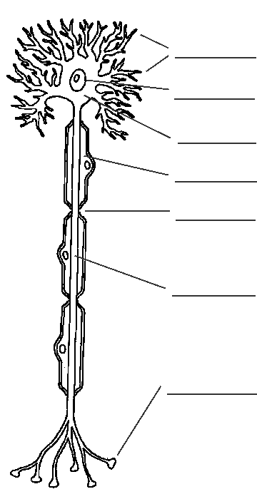
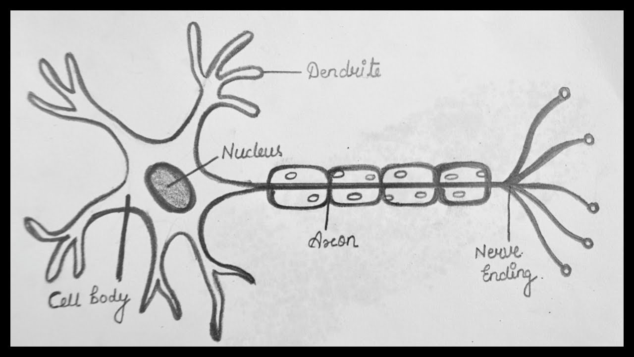
Diagram of neuron with labels. Neuron Labeling Worksheet - Nervous System Label The Neuron Label the parts of the neuron with the correct title: Consider this main learning worksheet to teach all about the fabulous neuron cells and the 6 parts. Label the central canal, grey matter, white matter, spinal nerve. Add the following labels to the diagram: Click on the dot and type in the correct structure. Neuron Diagram Unlabeled neuron, (1). axon, cell body, dendrites, nucleus, terminal. Unlabeled diagram of a motor neuron (try labeling: axon, dendrite, cell body, myelin, nodes of Ranvier, motor end plate).Read the definitions, then label the neuron diagram below. axon - the long extension of a neuron that carries nerve impulses away from the body of the cell. Sensory Neuron Diagram Illustrations & Vectors - Dreamstime Labeled diagram of the Neuron, nerve cell that is the main part of the nervous system. Abstract grey mesh background. Cells of human`s brain. Neuron and glial cells Microglia, astro. Cyte and oligodendrocyte. Vector diagram for educational, medical, biological and science use. medium.com › intuitive-deep-learning › autoencodersAutoencoders: Neural Networks for Unsupervised Learning Feb 18, 2019 · Supervised Learning deals with labelled data (e.g. an image and the label describing what is inside the picture) while Unsupervised Learning deals with unlabelled data (e.g. just the image itself ...
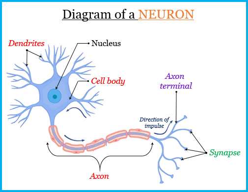
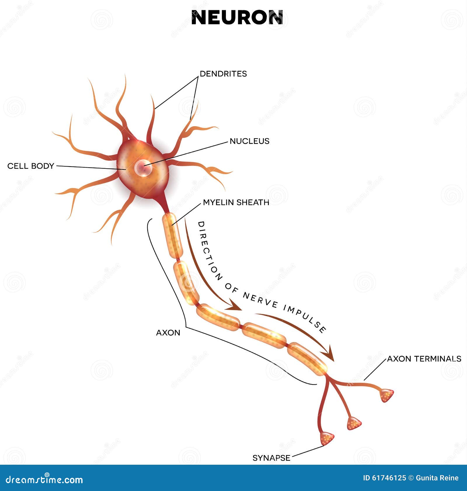
how to draw structure of neuron/neuron diagram labelled/diagram of ... Download this Premium Vector about Diagram of neuron anatomy, and discover more than 25 Million Professional Graphic Resources on Freepik. #freepik #vector #nervecell #nerve #neuron S Sharon Green Human body worksheets Human Nervous System Peripheral Nervous System Parasympathetic Nervous System Central Nervous System Three Types Of Neurons Labeled Diagram of the Neuron Stock Vector - Dreamstime Labeled diagram of the neuron, nerve cell that is the main part of the nervous system. neuron cell, sensory neuron, nervous system, main part, neuron, synapse, diagram, cell, system, nervous, nerve, labeled, vector, art, human, medical, science, drawing, health, chart, label, anatomy, body, isolated, cells, realistic More ID 61746125 Nervous System - Label the Neuron - TheInspiredInstructor.com Nervous System - Neuron: Nerve Cell. Choose the correct names for the parts of the neuron. (6) This neuron part receives messages from other neurons. (7) This neuron part sends on messages to other neurons. (8) This neuron part gives messages to muscle tissue. (9) This neuron part processes incoming messages. Labeled Diagram Of The Neuron, Nerve Cell That Is The Main Part ... - 123RF Illustration of Labeled diagram of the neuron, nerve cell that is the main part of the nervous system. vector art, clipart and stock vectors. Image 48129376. Discover millions of stock images, photos, video and audio.
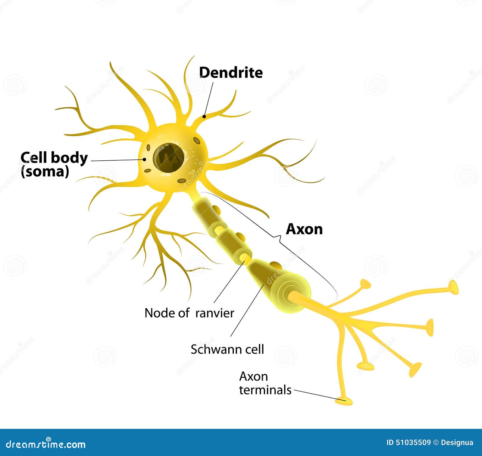
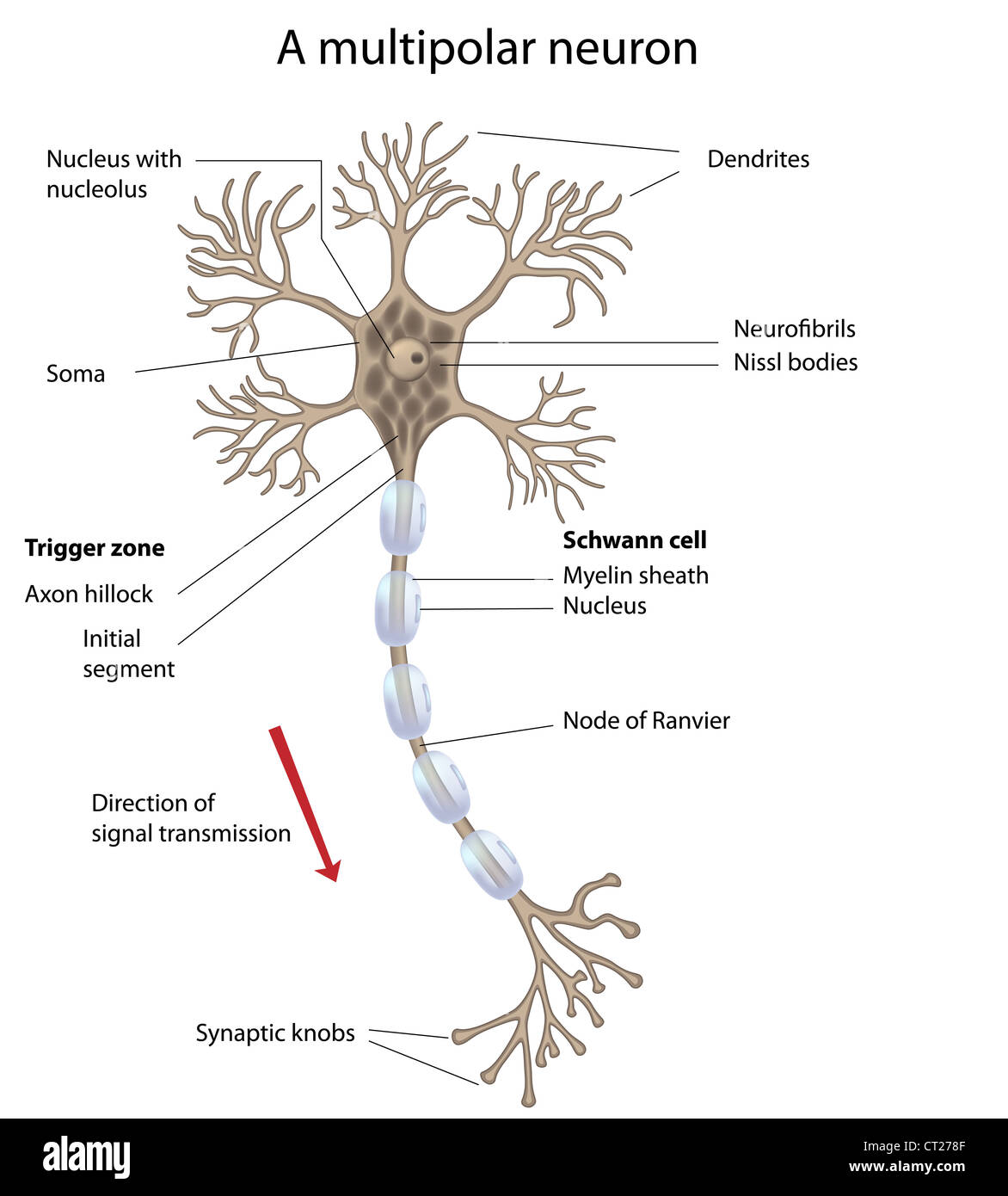
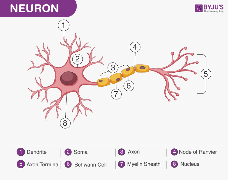
Label the Structures of the Neuron Diagram | Quizlet Label the Structures of the Neuron Diagram | Quizlet Label the Structures of the Neuron STUDY Learn Write Test PLAY Match + − Created by Nolan_Underwood53 Terms in this set (8) Cell Body ... Dendrites ... Axon Terminals/Synaptic Bulbs of other neurons ... Axon Hilock ... Schwann Cell ... Node of Ranvier ... Axon ... Actual Axon (Not Whole) ... Neuron under Microscope with Labeled Diagram - AnatomyLearner But, first, let's try to identify the following features from a neuron with the help of a labelled diagram. Cell body or perikaryon of a neuron Nucleus, cytoplasm, the plasma membrane of a neuron Nissl bodies in the cell body of a neuron An initial segment of axon and axon hillock Dendrites and axons of a neuron Axolemma and myelin sheath Draw a labelled diagram of neuron and label any four different parts ... Draw a labelled diagram of neuron and label any four different parts explain Here is the description of human neuron along with the diagram of the neuron and their parts. The neuron is a specialized and individual cell, which is also known as the nerve cell. A group of neurons forms a nerve. 1. Labeled diagram of the neuron stock illustration ... iStock Labeled Diagram Of The Neuron Stock Illustration - Download Image Now - Nerve Cell, Label, Labeling Download this Labeled Diagram Of The Neuron vector illustration now. And search more of iStock's library of royalty-free vector art that features Nerve Cell graphics available for quick and easy download. Product #: gm653408480 $ 12.00 iStock In stock
Label the parts of a typical neuron (1, 2, 3, 4, 5, 6) shown ...
Neuron Diagram || Diagram Of A Neuron || How To Draw A Neuron ... - YouTube Neuron Diagram || Diagram Of A Neuron || How To Draw A Neuron Step By Step For Beginners.
How To Draw A Neuron Step By Step For Beginners
Draw the diagram of neuron and label any two parts. - Toppr Ask Name the parts labelled a,b,c,d,e,f,g and h in the neuron drawn below. Draw a diagram of the human nerve cell. Justify its shape with regards to its function.

neuralNetworks
Label Neuron Anatomy Printout - EnchantedLearning.com Read the definitions, then label the neuron diagram below. axon - the long extension of a neuron that carries nerve impulses away from the body of the cell. axon terminals - the hair-like ends of the axon. cell body - the cell body of the neuron; it contains the nucleus (also called the soma)

Neurons (Nerve Cells) Structure, Function & Types - Simply ...
› ~kimscott › slides7. Artificial neural networks - Massachusetts Institute of ... Neuron Unit Synapse Connection Synaptic strength Weight Firing frequency Signals pass fromUnit output Table 1 (left): Corresponding terms from biological and artificial neural networks. Adapted from Adapted from Mehrotra, Mohan, & Ranka. Figure 1 (below): Schematic diagram of a standard neural network design. the input units
Neuroscience for Kids - Fill In #1
Neurons (With Diagram) - Biology Discussion Neurons (= Nerve Cells): A neuron is a structural and functional unit of the neural tissue and hence the neural system. Certain neurons may almost equal the length of body itself. Thus neurons with longer processes (projections) are the longest cells in the body. Human neural system has about 100 billion neurons.

Sketch of a neuron (1) cell nucleus, 2) dendrite and 3) axon ...
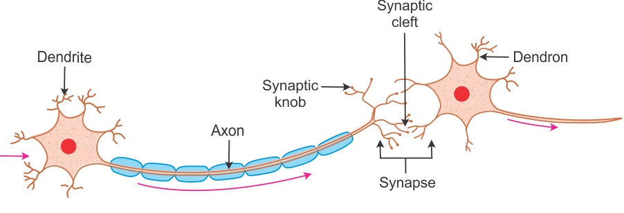
Neurons: Structure and Functions (With Diagram) - Psychology Discussion Each neuron consists of the nerve-cell and dendrites and axon. The dendrites look like the branches of a tree. The axon looks like a long slender thread without branches and ends in an end-brush. Synapse: The junction of two neurons is called the 'synapse'.

Well labelled diagram of neuron preferably hand drawn ...
byjus.com › biology › diagram-of-neuronA Labelled Diagram Of Neuron with Detailed Explanations - BYJUS A Labelled Diagram Of Neuron with Detailed Explanations Biology Biology Article Diagram Of Neuron Diagram Of Neuron A neuron is a specialized cell, primarily involved in transmitting information through electrical and chemical signals. They are found in the brain, spinal cord and the peripheral nerves. A neuron is also known as the nerve cell.

label the neuron - Clip Art Library
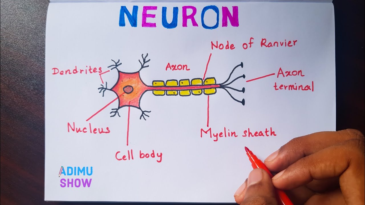
Neuron Diagram Labeled | EdrawMax Template In the following Neuron labeled diagram, we have dendrite, cell body, axon, myelin sheath, Schwann cell, a node of Ranvier, axon terminal, and nucleus. It should be noted here that a labeled diagram can be used in mathematics, science, and language arts to help the students or researchers understand the relationship among different parts of the diagram.

Schematic diagram of neuron structure | Download Scientific ...
neuron label and color - TeachersPayTeachers This is a 15 page PowerPoint slideshow on the basics of a neuron. It encourages the student to create a diagram, label, and color it. Then 5-6 links are provided where the student can practice on various neuron labeling websites. At the end is an edible and play doh model of the neuron to encourage further projects.

Neuron PowerPoint Diagram | Neurons, Neuron diagram, Diagram
Label Parts of a Neuron Diagram | Quizlet The chemical signals that neurons use to communicate with other neurons and cells. It is the chemical involved in impulse transmission. Neuron transportation. Neurons generally transport signals in one direction from the dendrites, through the soma, along the axon and unto the terminal buttons. Nodes of Ranvier.
A Guide to Understand Neuron with Neuron Diagram | EdrawMax ...
› articles › s41593/022/01041-5Single-neuron projectome of mouse prefrontal cortex | Nature ... Mar 31, 2022 · Top, a diagram and an example neuron with the over-represented pattern of projections to MOp, SSp, and SSs. Extended Data Fig. 6 Diversity of projections to lateral and central cortical subnetworks.

Neuron Structure Doodle Docs Graphic Organizer for Labeling Parts of a Neuron
Neuron Labelling Teaching Resources | Teachers Pay Teachers This reviews the structure, function and information processing done by the lobes and parts of the brain along with the parts of a nerve cell. Once this activity is loaded onto the students' devices they drag and drop the labels on to the appropriate places on diagrams. There are 12 structures to label on the brain and 6 to label on the neuron.
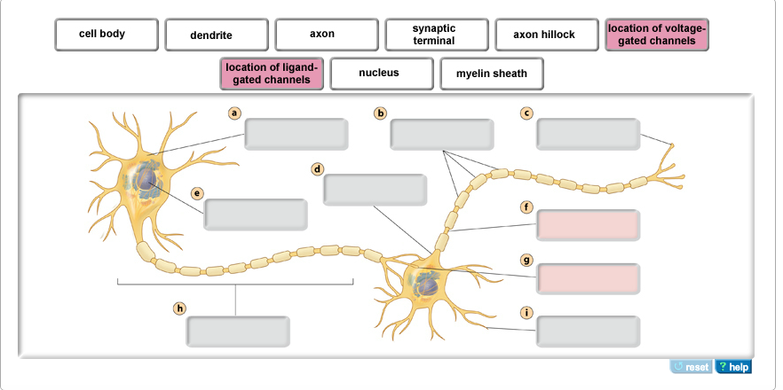
Solved Part A - Neuron structure Drag the labels to their ...
› photos › diagram-of-bodyDiagram Of Body Organs Female Pics Pictures, Images ... - iStock Mind Mapping Landscape Vibrant landscape vector illustration is showing a team using a diagram to visually organize information. Mind map is a graphic technique for visualizing connections between several ideas or pieces of information and it is represented as a central concept, and other ideas branch out of those (concept known as a spider diagram).

Neuron Diagram || Diagram Of A Neuron || How To Draw A Neuron Step By Step For Beginners
Draw a labelled diagram of a neuron. - toppr.com A myelinated nerve fibre is enveloped by Schwann cells, which form a myelin sheath around the axon. Reason : An unmyelinated nerve fibre is not enclosed by a Schwann cell and hence does not form a myelin sheath around the axon. Medium.
Neuron Diagram || Diagram Of A Neuron || How To Draw A Neuron Step By Step For Beginners
pressbooks.uwf.edu › medicalterminology › chapterNervous System – Medical Terminology for Healthcare Professions Labels read (top, left): pons, inferior olive, (top, right) cerebellum, deep cerebellar white matter (arbor vitae). In the top panel, a lateral view labels the location of the cerebellum and the deep cerebellar white matter. In the bottom panel, a photograph of a brain, with the cerebellum in pink is shown. [Return to Figure 8.7].
/548299803-56a796543df78cf7729765c8.jpg)
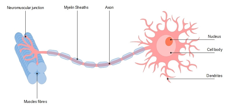
Parts of a Neuron and How Signals are Transmitted
Neuron Diagram & Types | Ask A Biologist - Arizona State University They pass signals from one neuron to the next inside the central nervous system. Pyramidal neurons are named after the shape of their cell body, which looks like a pyramid. They have one axon and two main dendrite branches. These cells pass signals inside the brain and tell your muscles to move.
Draw the structure of a neuron and label and following parts ...
Labeled Neuron Diagram | Science Trends Neurons are a type of cell and are the fundamental constituents of the nervous system and brain. Neurons take in stimuli and convert them to electrical and chemical signals that are sent to our brain. There are 3 major kinds of neurons in the spinal cord: sensory, motor, and interneurons.

Draw a well labelled diagram of neuron and label schwann cell. also mention the function of neur...
byjus.com › biology › skin-diagramSkin Diagram with Detailed Illustrations and Clear Labels - BYJUS Skin Diagram The largest organ in the human body is the skin, covering a total area of about 1.8 square meters. The skin is tasked with protecting our body from external elements as well as microbes.

Nerve Cell Labeled Diagram Stock Illustration | Adobe Stock
Diagram Quiz on Neuron Structure and Function (Labeling Quiz) Diagram Quiz on DNA replication. 1. Identify the cell type in the above figure. 2. In the figure, labeled '1' receives impulses from adjacent neuron. It is called the. 3. In the figure, labeled '2' is the short filaments from the cell body that carries impulses from dendrites to the cell body which is the. 4.

Label the parts of a neuron in the following diagram ...

ATS Science Neuron Label Diagram | Quizlet

Diagram Quiz on Neuron Structure and Function (Labeling Quiz)

File:Complete neuron cell diagram en.svg - Wikipedia

misscaityb I bet you could still label this! | Anatomy and ...

Diagram of Neuron Anatomy. Illustration of Neuron Anatomy ...
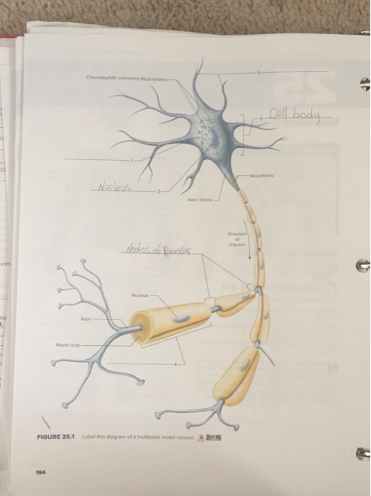
Solved ures Nucieus Axon FIQURE 25.1 Label this diagram of a ...

Neuron Label

Schematic Vector Diagram Neuron Nerve Cell Stock Vector ...

draw a labelled diagram of neuron and label any four ...
Motor Neuron, Detailed and Accurate, Labeled Stock Vector ...

Neuron Labeling Activity worksheet
Draw a labeled diagram of a neuron - Tutorix
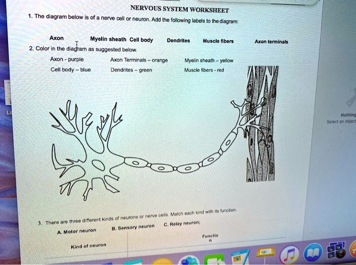
SOLVED: NERVOUS SYSTEM WORKSHEET nenve cell neuron. Add Ine ...
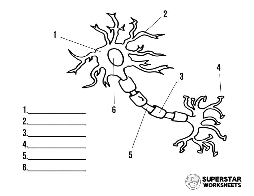
Neuron Cell Worksheets - Superstar Worksheets
Draw a diagram of neuron and name and label the part (a ...
Labeled Diagram of the Neuron Stock Vector - Illustration of ...

Solved Label the following diagram of a motor neuron. Each ...

draw a labelled diagram of a neuron j8t76t088 -Biology ...

Draw the diagram of neuron and label any two parts.

Neuron Label Diagram | Quizlet
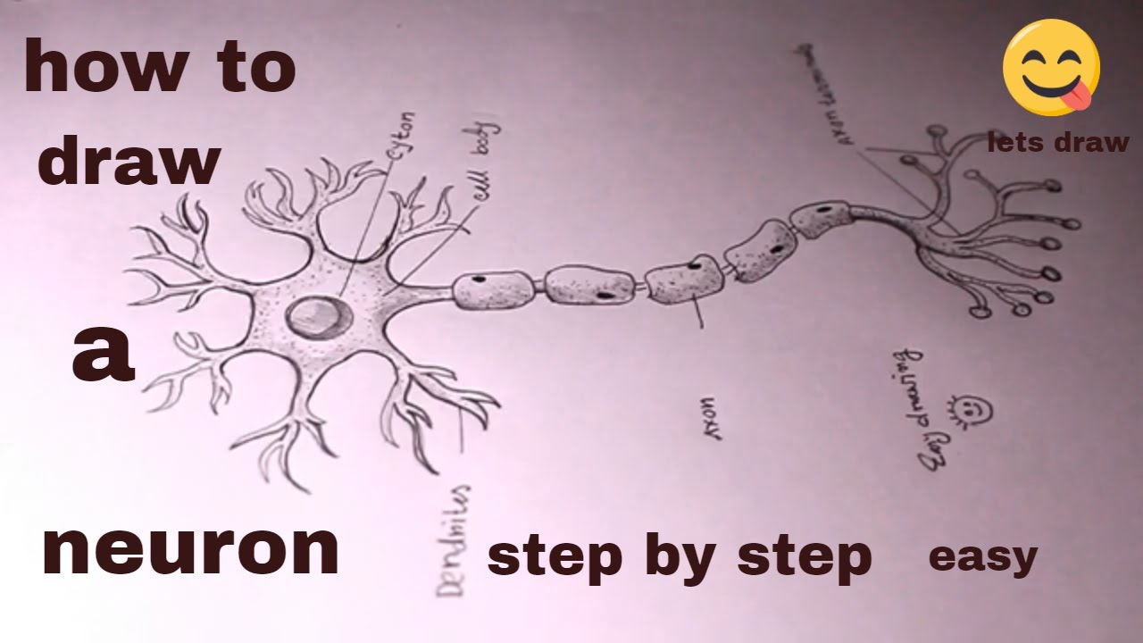
How TO Draw a neuron/draw neuron diagram/neuron drawing
Neuron diagram hi-res stock photography and images - Alamy

Biopsychology: Sensory, Relay and Motor Neurons | Psychology ...
A Labelled Diagram Of Neuron with Detailed Explanations
Post a Comment for "44 diagram of neuron with labels"