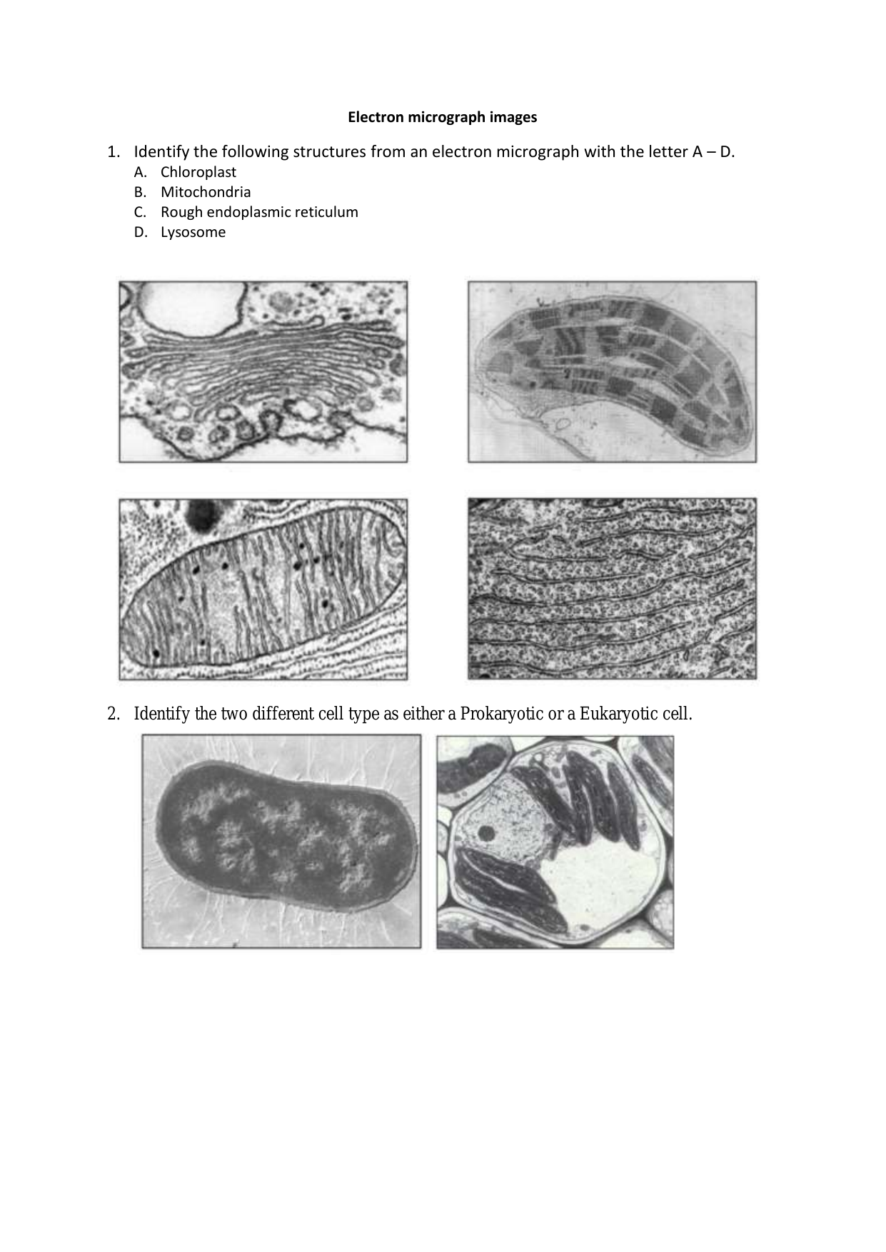
45 electron micrograph labeled
Scanning Electron Microscope (SEM) Scanning electron microscope is a classification of electron microscope that uses raster scanning to produce the images of a specimen by scanning using a focused electron beam on the surface of the specimen. An SEM creates magnified images of the specimen by probing along a rectangular area of the specimen with a focused electron beam. Nanogold-Antibody Conjugates - NANOPROBES This scanning transmission electron microscope (STEM) image clearly shows that labeling with Monomaleimido-Nanogold® (arrows) occurs specifically at a hinge thiol site on the IgG molecule. Develop further with our Silver Enhancers and Gold Enhancers for EM Light and Confocal Microscopy
Cathodoluminescence imaging of cellular structures labeled with ... Electron microscopy reveals cellular structures in incredible detail; however, the specific localization of nanoscale structures in its native, cryo-fixed environment is still in its infancy. The...
Electron micrograph labeled
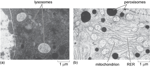
Electron Microscope- Definition, Principle, Types, Uses, Labeled Diagram An electron microscope is a microscope that uses a beam of accelerated electrons as a source of illumination. It is a special type of microscope having a high resolution of images, able to magnify objects in nanometres, which are formed by controlled use of electrons in a vacuum captured on a phosphorescent screen. Plant Cell: Meaning, Components, Structure, Functions & Parts - Embibe Despite the fact that plant and animal cells are both eukaryotic and share a few cell organelles, plant cells perform different roles than animal cells. When the cells are inspected under an electron microscope, some of these changes become obvious. In this article read more about Plant Cell, Diagram, Functions, and Types. Sperm Under Microscope with Labeled Diagram - AnatomyLearner The normal light microscope easily shows these stereocilia of the epididymal ducts. But, the electron microscope will show a clear view of these stereocilia. You will also see the agranular endoplasmic reticulum, lysosome, and prominent Golgi bodies in these lining epithelia of the epididymis (with an electron microscope).
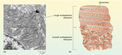
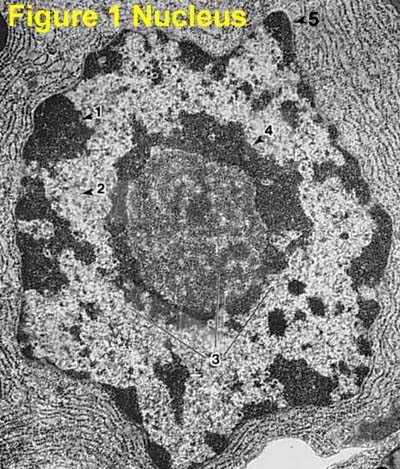
Electron micrograph labeled. 8.2: Transmission Electron Microscopy - Chemistry LibreTexts Transmission electron microscopy (TEM) is a form of microscopy which in which a beam of electrons transmits through an extremely thin specimen, and then interacts with the specimen when passing through it. The formation of images in a TEM can be explained by an optical electron beam diagram in Figure 8.2. 1. Scanning Electron Microscope (SEM)- Definition, Principle, Parts ... The first Scanning Electron Microscope was initially made by Mafred von Ardenne in 1937 with an aim to surpass the transmission electron Microscope. He used high-resolution power to scan a small raster using a beam of electrons that were focused on the raster. Parts of a microscope with functions and labeled diagram - Microbe Notes Parts of a microscope with functions and labeled diagram April 19, 2022 by Faith Mokobi Having been constructed in the 16th Century, Microscopes have revolutionalized science with their ability to magnify small objects such as microbial cells, producing images with definitive structures that are identifiable and characterizable. Label This Transmission Electron Micrograph / Microscopy Innovations ... The eluted conjugate is now ready for visualization by negative stain or cryo electron microscopy. Label the transmission electron micrograph of the nucleus. (d) a representative micrograph containing . Of machine learning to generalize metadata from a subset of labeled data,. In electron microscopy, however, true genetic encoded multilabeling.
Here's why people with allergic asthma are at lower COVID-19 risk Healthy human cells (labeled pink) from the lining of airways grow in "lawns" laced with some mucus (green) in this colorized electron micrograph. C. Ehre, C.B. Morrison et al/PNAS 2022 (CC BY ... 3D electron microscopy dataset reveals a zebrafish blood stem cell ... This allowed them to see and track the real-time development of a blood stem cell in the microenvironment of a live organism, then zoom in even further on the same cell using electron microscopy. "First, we identified single fluorescently labeled stem cells by light sheet or confocal microscopy," Tamplin says. "Next, we processed the same ... High throughput, label-free isolation of circulating tumor cell ... (Right) Scanning electron micrograph of a blood-spiked LNCaP cluster as captured by one of the wells on the device (see "Methods": SEM sample preparation and imaging). Scale bar, 20 μm. c Schematic... animal cell under electron microscope labelled - Be A Terrific Memoir ... Animal Cell Diagram Under Microscope Labeled. Here is an electron micrograph of an animal cell with the labels superimposed. An animal cell represents an eukaryotic cell in which true nucleus and other membrane-bound organelles such as mitochondria Golgi bodies and lysosomes are present. Function cell does in the body.
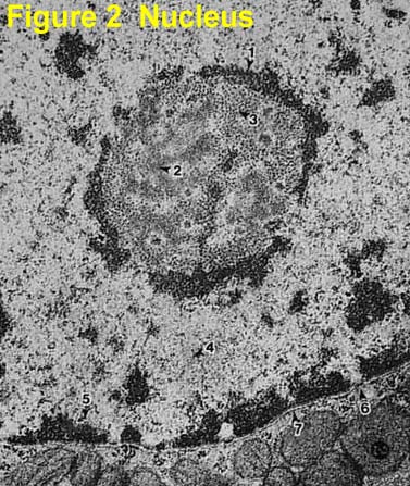
Label This Transmission Electron Micrograph - Kaiden Brown Provide the labels for the electron micrograph in figure 12.8. Label the transmission electron micrograph of the nucleus. Label the transmission electron micrograph of the nucleus. Transmission electron microscopy (tem) is a microscopy technique in which a beam of electrons is transmitted through a specimen to form an image. Light Microscope Labeled - how scanning electron microscopes work ... Light Microscope Labeled - 16 images - senior biology cell theory microscopy, what is a light microscope with pictures, 29 you will love labeling a compound microscope db, microscope imaging station gallery, Microscope Types (with labeled diagrams) and Functions The shorter wavelength of electrons compared to visible light photons helps the observer achieve a very high resolving power compared to normal microscopes thereby aiding observers to see very tiny objects clearly. Electron microscope labeled diagram The different types of electron microscopes are: Transmission Electron Microscope Electron Microscope Principle, Uses, Types and Images (Labeled Diagram ... Ans: An electron microscope has an evacuated column that is vacuum sealed and houses a cathode, anode, condenser magnet, scatter aperture, specimen chamber, objective lens, fluorescent screen, photographic plate and its transport machinery. This is the reason why the microscope is bulky in size. Q4.
7,218 Electron Micrograph Stock Photos, Pictures & Royalty ...
Transmission Electron Microscope (TEM)- Definition, Principle, Images Parts of a microscope with functions and labeled diagram Amazing 27 Things Under The Microscope With Diagrams Light Microscope- Definition, Principle, Types, Parts, Labeled Diagram, Magnification Scanning Electron Microscope (SEM)- Definition, Principle, Parts, Images Transmission Electron Microscope (TEM) Images

USMLE Pathology Slides — Cell structures, electron microscopy ...
Neuron under Microscope with Labeled Diagram - AnatomyLearner But, first, let's try to identify the following features from a neuron with the help of a labelled diagram. Cell body or perikaryon of a neuron Nucleus, cytoplasm, the plasma membrane of a neuron Nissl bodies in the cell body of a neuron An initial segment of axon and axon hillock Dendrites and axons of a neuron Axolemma and myelin sheath
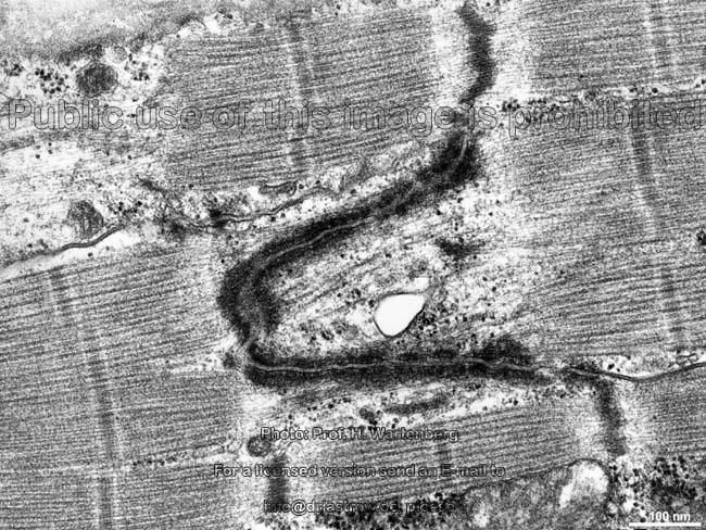
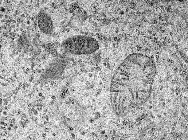
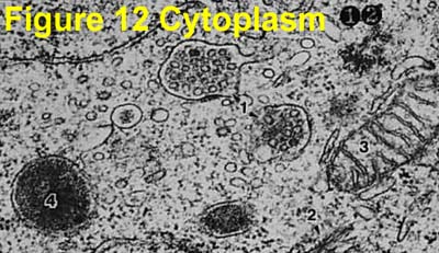
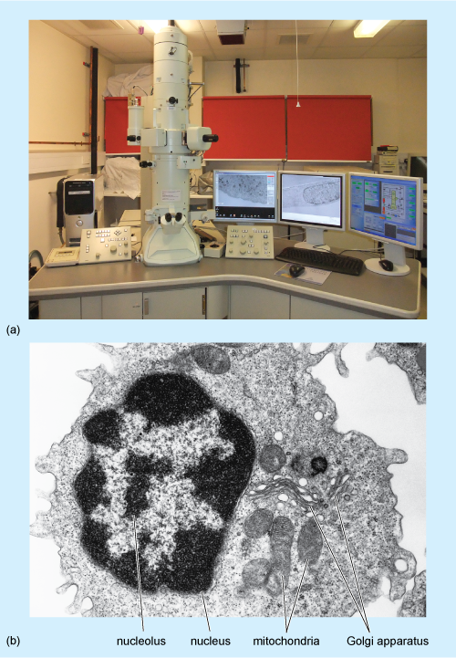
cell and organelles Dr.Jastrow's electron microscopic atlas
Sperm Under Microscope with Labeled Diagram - AnatomyLearner The normal light microscope easily shows these stereocilia of the epididymal ducts. But, the electron microscope will show a clear view of these stereocilia. You will also see the agranular endoplasmic reticulum, lysosome, and prominent Golgi bodies in these lining epithelia of the epididymis (with an electron microscope).
A tour of the cell: View as single page
Plant Cell: Meaning, Components, Structure, Functions & Parts - Embibe Despite the fact that plant and animal cells are both eukaryotic and share a few cell organelles, plant cells perform different roles than animal cells. When the cells are inspected under an electron microscope, some of these changes become obvious. In this article read more about Plant Cell, Diagram, Functions, and Types.
A tour of the cell: View as single page
Electron Microscope- Definition, Principle, Types, Uses, Labeled Diagram An electron microscope is a microscope that uses a beam of accelerated electrons as a source of illumination. It is a special type of microscope having a high resolution of images, able to magnify objects in nanometres, which are formed by controlled use of electrons in a vacuum captured on a phosphorescent screen.

Cell Micrographs | BioNinja
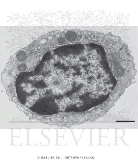
Electron Micrograph of a Lymphocyte

Electron micrograph of an autoradiograph of a 12-day feather ...

Cell Micrographs | BioNinja
Electron micrograph

Cell Micrographs | BioNinja
Electron Micrographs
Electron Micrographs
Electron Micrographs

Scanning electron micrograph of the control and Fe(TOB ...

1.2 Skill: Interpretation of electron micrographs - YouTube

7,218 Electron Micrograph Stock Photos, Pictures & Royalty ...

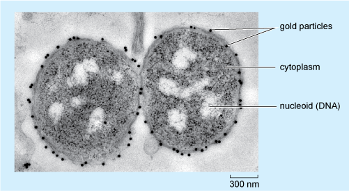
Electron micrographs of SPIO-labeled MSCs. A, Cell nucleus (N ...
A tour of the cell: View as single page
cell and organelles Dr.Jastrow's electron microscopic atlas
Electron Micrographs
Electron Micrographs
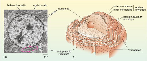
Level Labelling the organelles of a eukaryotic cell

2.2.3 Identify structures in electron micrographs of Ecoli ...
A and B) Electron micrograph of a cell labeled for/5-tubulin ...
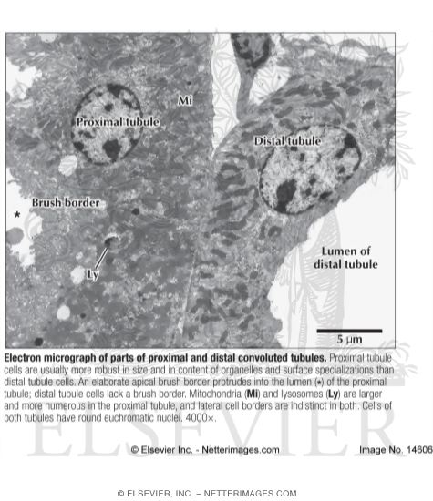
Electron Micrograph of Parts of Proximal and Distal ...
cell and organelles Dr.Jastrow's electron microscopic atlas

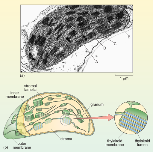
Electron micrograph of isolated chloroplasts with the major ...
cell and organelles Dr.Jastrow's electron microscopic atlas

Cell Micrograph Answers
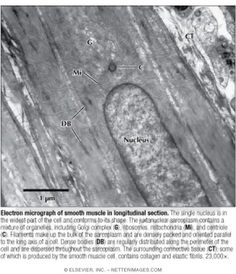
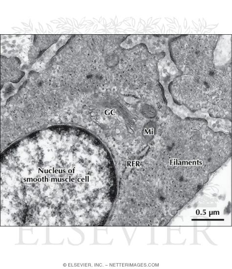
Electron Micrograph of Smooth Muscle In Longitudinal Section

Low-power electron micrograph of a TH+ perikaryon in tissue ...
A tour of the cell: View as single page
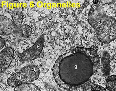
Electron Micrograph of Cell Organelles
A tour of the cell: View as single page
A tour of the cell: View as single page
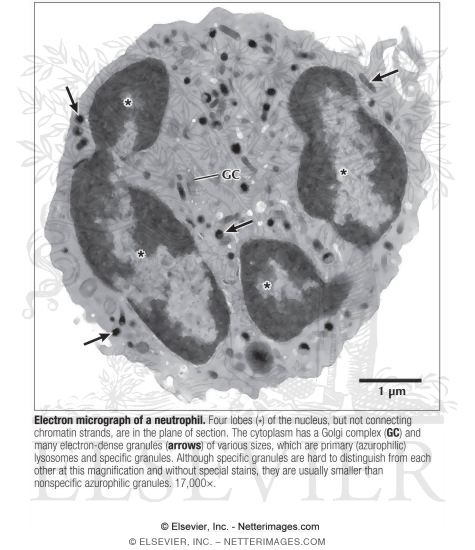
Electron Micrograph of a Neutrophil
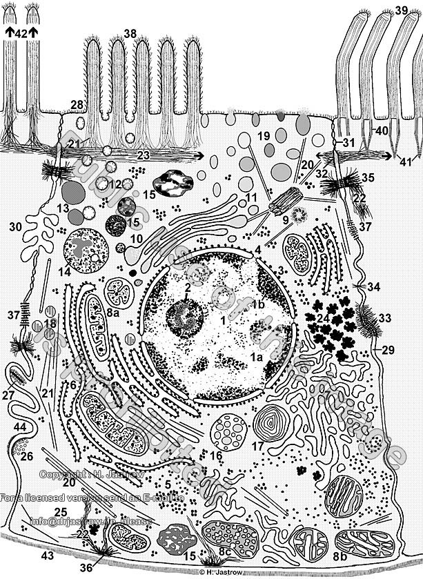
cell and organelles Dr.Jastrow's electron microscopic atlas
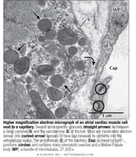
Higher Magnification Electron Micrograph of an Atrial Cardiac ...
Electron Micrograph of Actin and Intermediate Filaments In ...

2.3.3 Identify structures from electron micrographs of liver ...
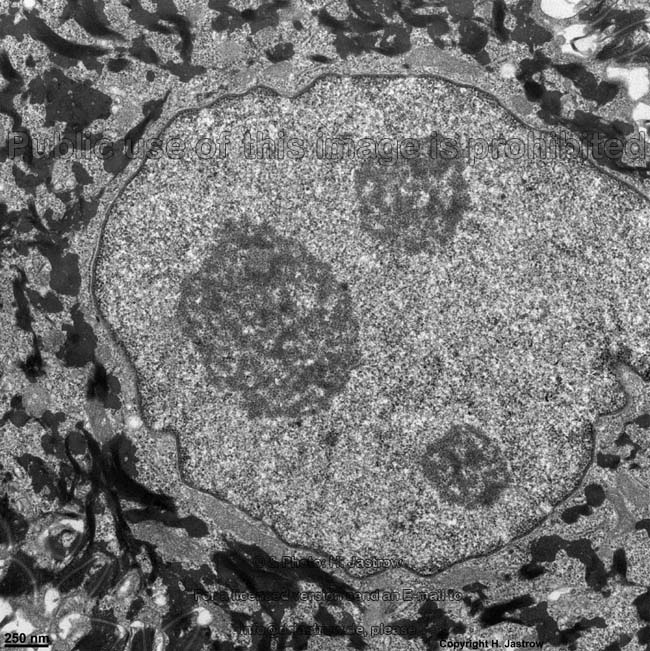
cell and organelles Dr.Jastrow's electron microscopic atlas

Electron Micrograph of Plasma Cells In Connective Tissue

7,218 Electron Micrograph Stock Photos, Pictures & Royalty ...

Transmission Electron Micrograph of transfected HL-1 cells ...
Post a Comment for "45 electron micrograph labeled"