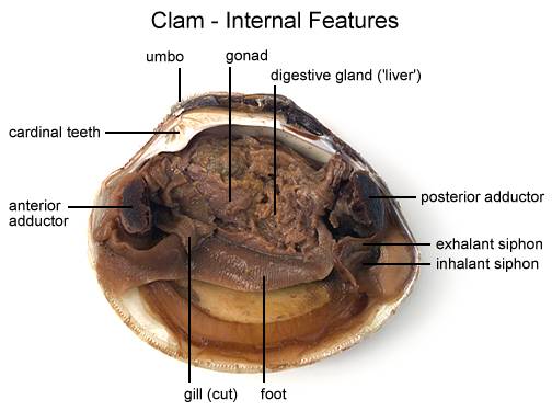
45 internal structures of a clam
CDC director announces shake-up, citing COVID mistakes Aug 18, 2022 · Walensky, who became director in January 2021, has long said the agency has to move faster and communicate better. Read more at Boston.com. Clam Dissection - BIOLOGY JUNCTION Clams are marine mollusks with two valves or shells. Like all mollusks, a clam has a mantle which surrounds its soft body. It also has a muscular foot which enables the clam to burrow itself in mud or sand. The soft tissue above the foot is called the visceral mass and contains the clam's body organs.
Clam Dissection Clams are marine mollusks with two valves or shells. Like all mollusks, a clam has a mantle which surrounds its soft body. It also has a muscular foot which enables the clam to burrow itself in mud or sand. The soft tissue above the foot is called the visceral mass and contains the clam's body organs. Taxonomy Kingdom - Animalia Phylum - Mollusca

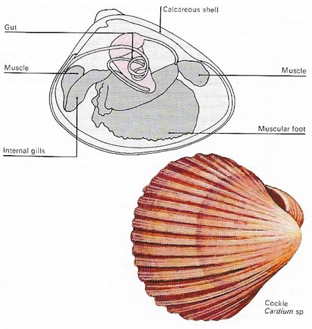
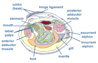
Internal structures of a clam
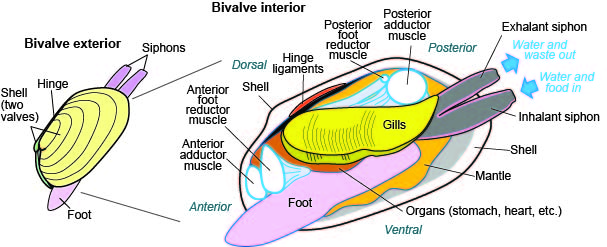
Bivalve shell - Wikipedia The anterior or front of the shell is where the byssus and foot are located (if the animal has these structures) and the posterior or back of the shell is where the siphon is located (again, if present— the scallops, for example, do not have siphons). Without being able to view these organs, however, determining anterior and posterior can be ... PDF Clam Dissection Information Sheet - Internal Anatomy A. Feeding Mechanism of clam. Food in mucous string Water enters the mantle cavity from the rear and is pulled forward by the beating of cilia to the gills and mouth. Water flowing over the gill is filtered, tiny food partic es External are caught in the mucus coating and carried by cilia, gill in a mucus string, to the mouth. Sand and other ... Clam study: the shell, the internal anatomy and how they feed Clam study: the shell, the internal anatomy and how they feed. Summary. Compare different sizes of shells and learn about how shells grow. Dissect a clam and discover that inside a familiar clam shell, often seen on the beach, there is a living animal. Identify the major body parts of a clam, and compare their function to equivalent organs in ...
Internal structures of a clam. PDF Investigation #5 - Clam Anatomy - COSEE The smallest clam is _____ cm wide and _____ cm long. The average length of all clams measured by the class is _____ cm Inside of the clam: 1). The thin, whitish flesh lining is called the _____. The mantle encloses all the internal organs of the clam. It is also where new shell is made as the clam grows. The internal, soft tissue anatomy of clam - ResearchGate Mantle: The soft parts of the clam tissues are are covered by the mantle, which is composed of two thin sheaths of tissue, thickened at the edges ( Fig.3) . The two halves of the mantle are... PDF Anatomy of a Clam - University of Florida This has a single, "limpet-like" shell on top, which is made of proteins and chitin reinforced with calcium carbonate, and is secreted by a mantle that covers the whole upper surface. The underside of the animal consists of a single muscular "foot". Clam - Wikipedia A clam's shell consists of two (usually equal) valves, which are connected by a hinge joint and a ligament that can be internal or external. The ligament provides tension to bring the valves apart, while one or two adductor muscles can contract to close the valves. Clams also have kidneys, a heart, a mouth, a stomach, and a nervous system.
ESL tongue twisters for English pronunciation practice Download English Video slide lessons for use on ipods, PCs or laptops.These videos are lesson presentations built with powerpoint. Learn & teach new vocabulary, pronunciation, spelling and sentence structures with videos that can be used for self-tutoring or teaching in larger classrooms. Internal Structure of the Clam 09 2 - YouTube Here is the second part to the Clam dissection for Tim Revell's Bio 2 class Spring 2009. Clam Diagram & Parts | What Is a Clam? | Study.com Below represents a diagram of the external and internal anatomy of a clam with some of the anatomy labeled as follows: a: adductor muscles ; b: gills ; h: heart ; o: mouth ; p: labial palps ; f ... Solved 3. Describe what it means for an organism to be | Chegg.com What structures of the internal anatomy of a clam might lead you to conclude that a clam is triploblastic? Expert Answer 100% (5 ratings) Ans) An organism can be called triploblastic when it has three germ layers namely ectoderm, mesoderm and endoderm. Generally, multicellular organisms are only triploblastic and they are bilatera …
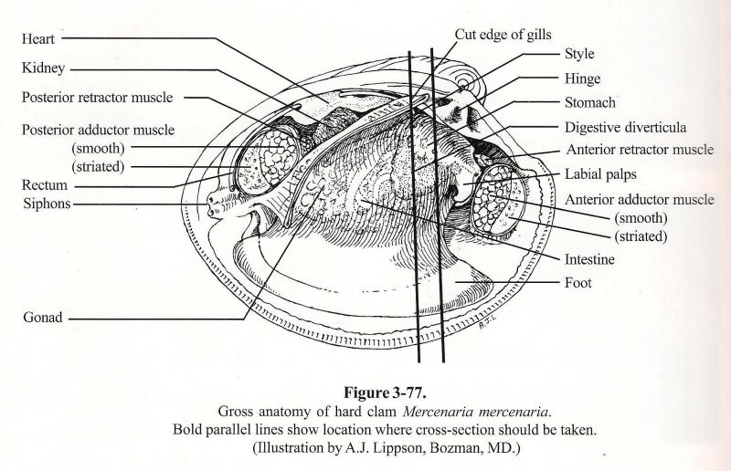
Clam anatomy and what their organs do by Ethan Enfinger - Prezi The vital Organs. The most important out of all the organs in the clam is the heart which pumps blood to all the other organs allowing them to live. Their is also the nervous system which tell parts of the clam to do certain things. Solved Results 1. Draw a diagram and label the internal - Chegg Results 1. Draw a diagram and label the internal anatomy of the clam. Which structures were difficult to locate? 2. In what ways does the clam differ from other mollusks? What modifications are critical for its unique habitat and lifestyle? Question: Results 1. Draw a diagram and label the internal anatomy of the clam. The hatchery culture of bivalves: a practical manual In clams and mussels the two adductor muscles are located near the anterior and posterior margins of the shell valves. The large, single muscle is centrally located in oysters and scallops. The muscle(s) close the valves and act in opposition to the ligament and resilium, which spring the valves open when the muscles relax. PDF Taxonomy, Anatomy, and Biology of the Hard Clam Internal Clam 1 Mantle Shell Anatomy • Covers visceral or body mass • Holds in fluid • Secrets new shell 2. Ant. adductor muscle 3. Post adductor musclePost. adductor muscle • Hold valves shut 4. Pericardium cavity • Region covered with thin Region covered with thin, dark membrane • Contains 2-chambered heart and kidney in a fluid-filled sac 5.
Anatomy – GL2019 Bivalvia
Clams Anatomy - Barnegat Bay Shellfish The shell is "spring loaded open" by the hinge. When alive, the two adductor muscles keep it tightly closed. Clam shells are made up in three layers: Nacreous Layer. Prismatic Layer. Periostracum Layer. Nacreous Layer (nacre) The innermost layer of the shell which touches the clam and connects it to the shell.

Review Of Clam Internal Anatomy 2022 - PeepsBurgh
anatomy of clam - Microsoft clam diagram labeled internal structures anatomy01 label incurrent excurrent siphon lab diagrams answer. Images For BIO 122 Lab klemow.wilkes.edu. unlabeled dissected lab earthworm labeled anterior wilkes dorsal bio. Clam Anatomy (Function Quiz) . clam. Leevonk.com leevonk.com. anatomy clam biology. LLA BIOLOGY: Squid Anatomy
Elements of biology, with special Elements of biology, with ...
internal anatomy of a clam Clam Dissection . dissection gills clam figure. Sea Cucumber Anatomy Dissection thebestsea.blogspot.com. dissection blowes asterias rubens. Internal Earthworm Anatomy . earthworm anatomy internal dissection earthworms septum ganglion faculty sdmiramar edu. Clam (Mollusca) Dissection - YouTube www ...
MARINE BIOLOGY: CLAM DISSECTION
Anatomy of the Geoduck Clam - Fisheries and Oceans Canada Anatomy of the Geoduck Clam. The general anatomy of geoduck clams was established from cultured juveniles and both normal and abnormal wild adults. The cultured geoduck clams (6 - 25 mm in shell length) were collected from ocean nursery sites in Georgia Strait, B.C. Adult clams were gathered from the east and west coast of Vancouver Island and ...
Biology:Clam/Freshwater Mussel Pre-lab Dissection
anatomy of clam Clam Internal Anatomy - Anatomy Drawing Diagram sen842cova.blogspot.com. clam anatomy dissection bivalve adductor clams bivalvia. CLAM IMAGES: Biology 201 people.stfx.ca. clam biology clams stfx. Clam Anatomy - YouTube . Clam Dissection | Flickr - Photo Sharing! .

Lab 15 - Animal Diversity Intro - Clams (Part 1 of 5) - Kathy ...
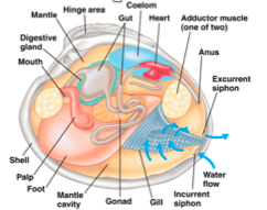
Clam structures and functions Flashcards | Quizlet gills. remove oxygen and food particles from the water; moves water throughout the clam using cilia to create a current through the siphons. palps. guide food particles into the mouth from the gills. esophagus. food travels through in order to be digested and sent to the stomach. stomach. stores food; digests to diffuse through the visceral mass.

mussel internal anatomy Diagram | Quizlet
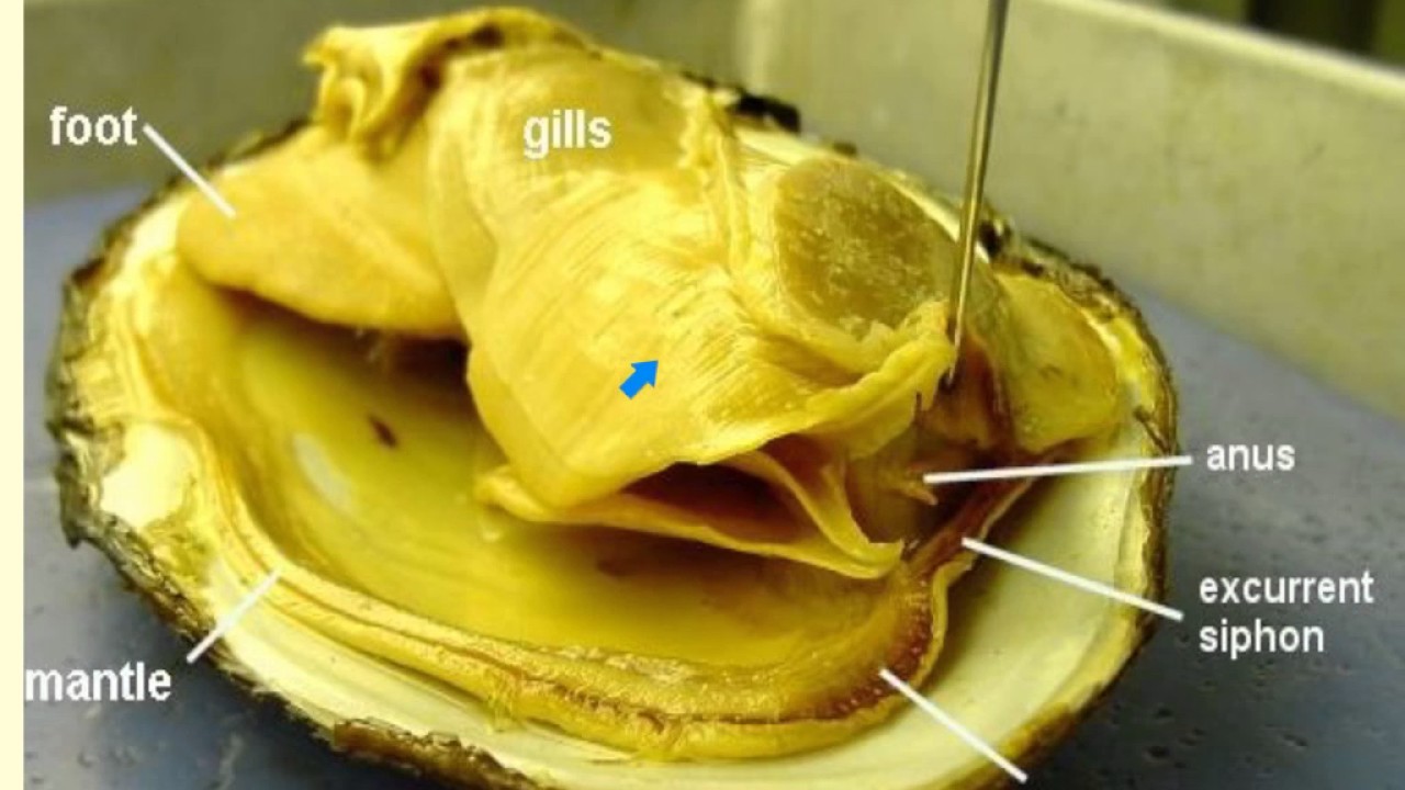
PDF Mussel and Clam Anatomy - Mt. San Antonio College Internal Anatomy of the Clam Gills Foot Mantle Labial palps . Title: Slide 1 Author: Sherry Created Date: 5/3/2012 11:11:29 AM ...

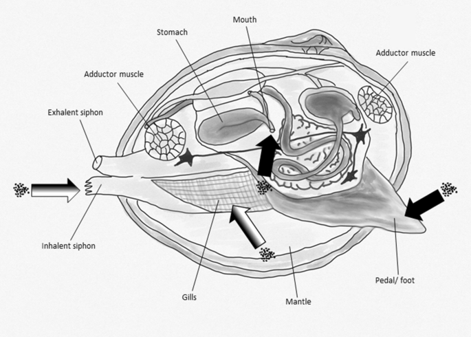
4 Internal anatomy of a typical heterodont bivalve. In this ...
Internal Structure of the Clam 09 1 - YouTube Here is the first part of the Clam dissection for Tim Revell's Bio 2 class 2009.
Lab 5: Phylum Mollusca
clam | mollusk | Britannica True clams, in the strict sense, are bivalves with equal shells closed by two adductor muscles situated at opposite ends of the shell, and with a powerful, muscular, burrowing foot. Clams characteristically lie buried from just beneath the surface to depths of about 0.6 metre (2 feet). They rarely travel over the bottom as do some other bivalves.

Clam Body Parts Discount, 57% OFF | www.vicentevilasl.com
Tiny oysters play big role in stabilizing eroding shorelines Aug 28, 2022 · Baby oysters, called spat, attached to a clam shell shortly before it was to be placed into the water in Beach Haven, N.J. on Aug. 19, 2022, as part of a shoreline stabilization plan using oyster ...
Clam Body Parts Sale, 58% OFF | www.cartenztactical.com
internal anatomy of a clam clam clams anatomy body foot which visceral mass giant organ siphon gonad incurrent reproductive male excurrent marine digestion animals female. Brain Anatomy On Mri boundbobskryptis.blogspot.com. brain anatomy mri radiology nuclei basal ganglia capsule internal assistant interna neuroanatomy neuroanatomia grey subthalamic radiologyassistant nl ...
bivalve
Clam Dissection Lab: Explained | SchoolWorkHelper Like all mollusks, a clam has a mantle which surrounds its soft body. It also has a muscular foot which enables the clam to burrow itself in mud or sand. The soft tissue above the foot is called the visceral mass and contains the clam's body organs. Objective To study the internal and external anatomy of a bivalve mollusk.

Internal Structure of the Clam 09 1
Clam Anatomy - Clams Ahoy The common hardshell clam Mercenaria mercenaria, better known as a cherrystone, has a mouth, labial palps (antecedents of lips), a stomach, separate digestive gland, an intestine, nerve cord and an anus. The foot of a clam is a curved flesh protrusion from the perimeter of the anterior flesh.
GIANT CLAM PRODUCTION IN THE REPUBLIC OF THE MARSHALL ISLANDS ...
Clam study: the shell, the internal anatomy and how they feed Clam study: the shell, the internal anatomy and how they feed. Summary. Compare different sizes of shells and learn about how shells grow. Dissect a clam and discover that inside a familiar clam shell, often seen on the beach, there is a living animal. Identify the major body parts of a clam, and compare their function to equivalent organs in ...
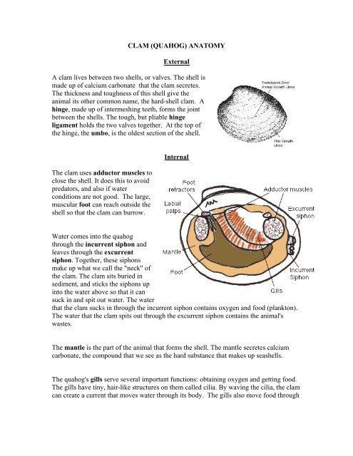
CLAM (QUAHOG) ANATOMY
PDF Clam Dissection Information Sheet - Internal Anatomy A. Feeding Mechanism of clam. Food in mucous string Water enters the mantle cavity from the rear and is pulled forward by the beating of cilia to the gills and mouth. Water flowing over the gill is filtered, tiny food partic es External are caught in the mucus coating and carried by cilia, gill in a mucus string, to the mouth. Sand and other ...

Microstructure and mechanical property of Ruditapes ...
Bivalve shell - Wikipedia The anterior or front of the shell is where the byssus and foot are located (if the animal has these structures) and the posterior or back of the shell is where the siphon is located (again, if present— the scallops, for example, do not have siphons). Without being able to view these organs, however, determining anterior and posterior can be ...

Clam Dissection Lab

Clams - the muscular system

Lab 15 - Animal Diversity Intro - Clams (Part 1 of 5) - Kathy ...

New Page 1
UCSC Biology 136 M/Vtext

Clam Dissection - Mrs. Skakal's BVW Webpage

clam internal anatomy Diagram | Quizlet
clam anatomy
Bioaccumulation assessment of nanomaterials using freshwater ...

Science Magazine: EXTERNAL AND INTERNAL FEATURES OF A CLAM
Name_________________________ THE CLAM
chapter 35 Flashcards | Chegg.com
Clam Dissection Information Sheet – Internal Anatomy
Shellfish | Roger Williams University
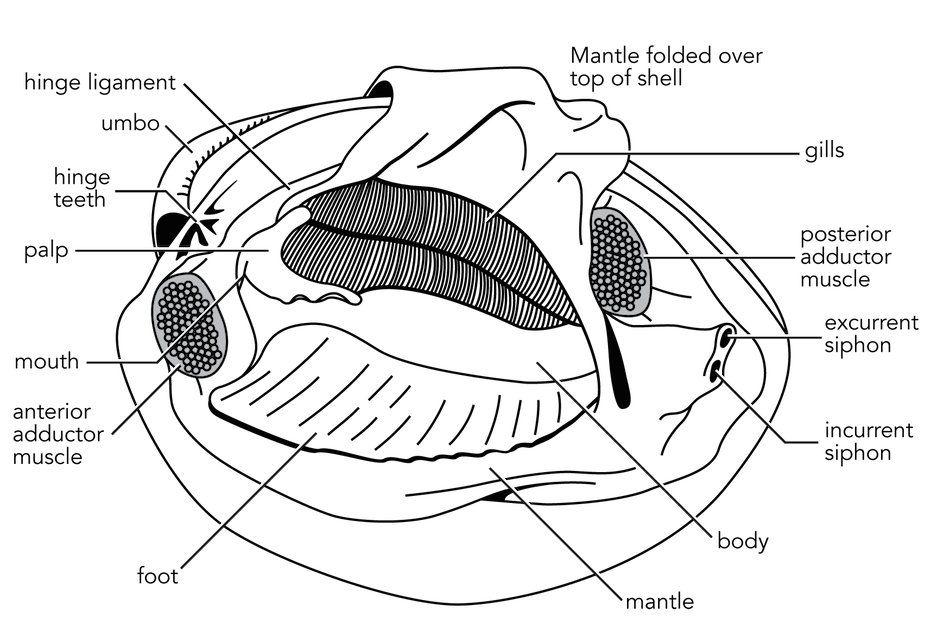
Fig3.63-InternalStructureClam.png

Clam Project

Clam Internal Anatomy Quiz

Clam Dissection - BIOLOGY JUNCTION
CLAM DISSECTION HANDOUT Fig 6-6a Fig 6-6b
Solved] Draw a diagram and label the internal anatomy of the ...
Zoology Animal Morphology Internal Anatomy Example Stock ...

The hatchery culture of bivalves: a practical manual

Mollusca | Veterian Key

The hatchery culture of bivalves: a practical manual

Henderson State University Zoology Mollusc Quiz
Hard clams - Barnegat Bay

The internal, soft tissue anatomy of clam | Download ...
Bivalves (pelecypods, clams, etc.), Fossils, Kentucky ...
Clam Dissection - BIOLOGY JUNCTION
Post a Comment for "45 internal structures of a clam"